 뒤로
뒤로Nucleic Acids: Structure, Function, and Genetic Implications
스터디 가이드 - 스마트 노트
 자료에 맞춘 맞춤형 노트, 핵심 정의, 예시, 맥락을 확장해 제공합니다.
Nucleic Acids in Biochemistry
Introduction to Nucleic Acids
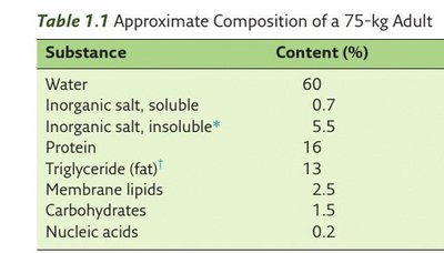
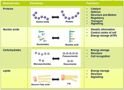
Nucleic acids are essential biomolecules that control cellular activities by storing and transmitting genetic information. They are the least abundant biomolecules in the human body but are vital for the storage of heritable information and the regulation of protein production.
Definition: Nucleic acids are unbranched polymers composed of repeating nucleotide monomers.
Main Types: Deoxyribonucleic acid (DNA) and Ribonucleic acid (RNA).
Functions: Storage and transmission of genetic information, and regulation of protein synthesis.
Cellular Abundance and Role
Nucleic acids, though present in small amounts compared to other biomolecules, play a central role in cellular function by dictating the synthesis of proteins, which are responsible for most cellular processes.
Types and Structure of Nucleic Acids
DNA and RNA: Structure and Function
There are two main types of nucleic acids found in cells:
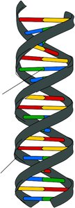
DNA (Deoxyribonucleic Acid): Double-stranded polynucleotide that stores genetic information and is located in the nucleus.
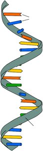
RNA (Ribonucleic Acid): Single-stranded polynucleotide that translates genetic information from DNA into proteins. Synthesized in the nucleus and functions in the cytoplasm.
Nucleotides: Building Blocks of Nucleic Acids
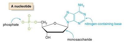
Nucleotides are the monomers of nucleic acids, each consisting of three components:
Monosaccharide: Deoxyribose in DNA, ribose in RNA.
Nitrogen-containing base: Purines (adenine, guanine) and pyrimidines (cytosine, thymine in DNA; uracil in RNA).
Phosphate group
DNA Structure and Organization
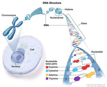
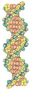
DNA is organized into chromosomes within the nucleus, where it is tightly wrapped around histone proteins to save space. The double helix structure consists of two antiparallel strands held together by complementary base pairing.
Base Pairing: Adenine (A) pairs with Thymine (T) via two hydrogen bonds; Guanine (G) pairs with Cytosine (C) via three hydrogen bonds.
Antiparallel Strands: One strand runs 5' to 3', the other 3' to 5'.
DNA Synthesis and Directionality
DNA synthesis involves the formation of phosphodiester bonds between nucleotides, catalyzed by DNA polymerase. The process requires high-energy deoxynucleotide triphosphates (dNTPs), which provide the energy for polymerization.
5' and 3' Ends: The 5' end has a free phosphate group; the 3' end has a free hydroxyl group. DNA is synthesized and read from 5' to 3'.
Complementary Base Pairing: Ensures accurate replication and transcription.
Central Dogma of Molecular Biology
Flow of Genetic Information
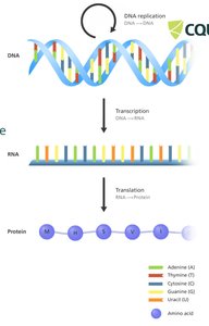
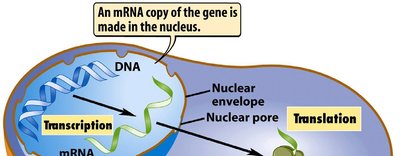
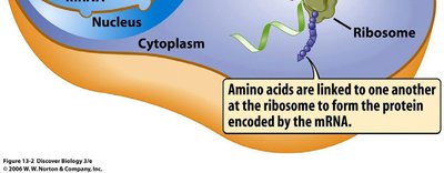
The central dogma describes the flow of genetic information from DNA to RNA to protein. Genes in DNA are transcribed into messenger RNA (mRNA), which is then translated into proteins by ribosomes in the cytoplasm.
Transcription: DNA → RNA
Translation: RNA → Protein
Historical Context: Discovery of DNA Structure
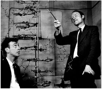
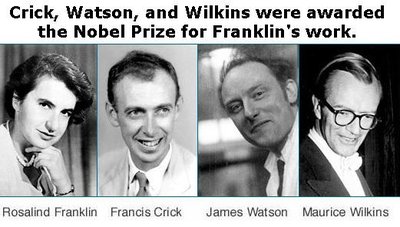
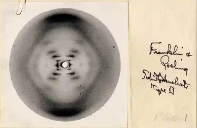
The double helix structure of DNA was elucidated in 1953 by Watson and Crick, with critical contributions from Rosalind Franklin's X-ray crystallography data. Franklin's "Photo 51" provided key evidence for the helical structure, though her contributions were not fully recognized during her lifetime.
High-Energy Nucleotides: ATP
Structure and Function of ATP
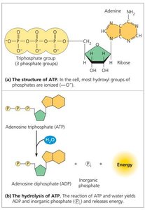
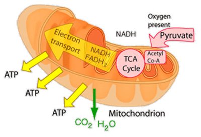
Adenosine 5'-triphosphate (ATP) is a high-energy nucleotide that stores energy in its phosphate bonds. Hydrolysis of ATP releases energy required for various cellular processes. ATP is synthesized in mitochondria and used throughout the cell.
Structure: Adenine base, ribose sugar, and three phosphate groups.
Function: Universal energy currency of the cell.
Transcription: Production of RNA from DNA
Process of Transcription
Transcription is the synthesis of RNA from a DNA template. The DNA double helix unwinds, and RNA polymerase synthesizes a complementary RNA strand using one DNA strand as a template. The resulting RNA is single-stranded and contains uracil (U) instead of thymine (T).
Base Pairing in Transcription: Adenine pairs with Uracil (A-U); Cytosine pairs with Guanine (C-G).
Direction: RNA is synthesized from 5' to 3'.
Types of RNA
There are several types of RNA, each with specific roles in protein synthesis:
mRNA (Messenger RNA): Carries genetic information from DNA to ribosomes.
tRNA (Transfer RNA): Brings amino acids to the ribosome during translation.
rRNA (Ribosomal RNA): Structural and catalytic component of ribosomes.
Translation: Protein Synthesis
Genetic Code and Translation
The genetic code consists of codons—triplets of nucleotides on mRNA—that specify amino acids. Translation occurs in the ribosome, where tRNA molecules match their anticodons to mRNA codons and add the corresponding amino acids to the growing polypeptide chain.
Start Codon: AUG (codes for Methionine)
Stop Codons: UAA, UAG, UGA (signal termination of translation)
Genetic Mutations and Diseases
Types of Genetic Mutations
Mutations are changes in the DNA sequence that can affect protein structure and function. Major types include:
Silent Mutation: No change in amino acid sequence due to redundancy in the genetic code.
Missense Mutation: Alters a single amino acid in the protein.
Nonsense Mutation: Introduces a premature stop codon, truncating the protein.
Frameshift Mutation: Insertion or deletion of nucleotides shifts the reading frame, altering downstream amino acids.
Examples of Genetic Diseases
Early-Onset Familial Alzheimer's Disease: Caused by missense mutations in the APP gene, leading to toxic amyloid beta peptide accumulation.
Cystic Fibrosis: Caused by nonsense mutations in the CFTR gene, resulting in nonfunctional chloride channels and thick mucus production.
Tay-Sachs Disease: Caused by frameshift mutations in the HEXA gene, leading to accumulation of GM2 ganglioside and neuronal damage.
Summary Table: Key Biomolecules in the Human Body
Substance | Content (%) |
|---|---|
Water | 60 |
Inorganic salt, soluble | 0.7 |
Inorganic salt, insoluble | 5.5 |
Protein | 16 |
Triglyceride (fat) | 13 |
Membrane lipids | 2.5 |
Carbohydrates | 1.5 |
Nucleic acids | 0.2 |
