뒤로
뒤로Protein Analysis Techniques and Protein Structure: Study Notes for Biochemistry
스터디 가이드 - 스마트 노트
 자료에 맞춘 맞춤형 노트, 핵심 정의, 예시, 맥락을 확장해 제공합니다.
Protein Analysis Techniques
Introduction to Protein Separation and Purification
Understanding protein structure and function requires pure protein samples. Cells contain thousands of proteins, necessitating effective separation and purification methods based on properties such as size, charge, and binding affinity.
Classical methods: Utilize differences in protein size, charge, and binding properties.
Modern methods: Include DNA cloning and genome sequencing for protein production and identification.
Steps in Protein Purification
Source: Proteins are extracted from tissues or microbial cells.
Crude extract: Cells are lysed to release proteins into solution.
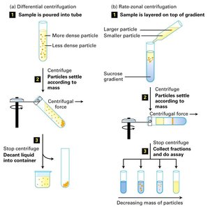
Differential centrifugation: Used to separate cellular components by mass and density.
Fractionation: Proteins are separated into fractions based on properties such as solubility, size, or charge.
Salting out: Protein solubility decreases at high salt concentrations, commonly using ammonium sulfate ((NH4)2SO4).
Dialysis
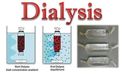
Dialysis separates proteins from small molecules by exploiting the size difference. The protein solution is placed in a semipermeable membrane bag, allowing small molecules to diffuse out while retaining proteins.
Column Chromatography
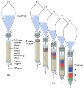
Column chromatography is a central technique for protein purification, separating proteins based on charge, size, or binding affinity. The stationary phase is a solid matrix, and the mobile phase is a buffered solution.
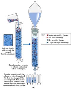
Ion-exchange chromatography: Separates proteins by net charge at a given pH using charged resins (cation or anion exchangers).
Size-exclusion (gel filtration) chromatography: Separates proteins by size; large proteins elute first.
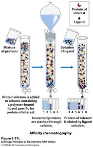
Affinity chromatography: Separates proteins based on specific binding to a ligand attached to the matrix.
Ion-Exchange Chromatography
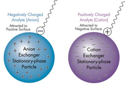
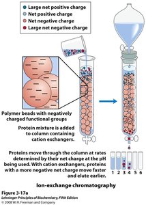
Proteins are separated based on their net charge. Cation exchangers have negatively charged groups and bind positively charged proteins, while anion exchangers have positively charged groups and bind negatively charged proteins.
Size-Exclusion (Gel Filtration) Chromatography
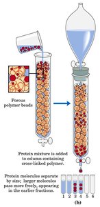
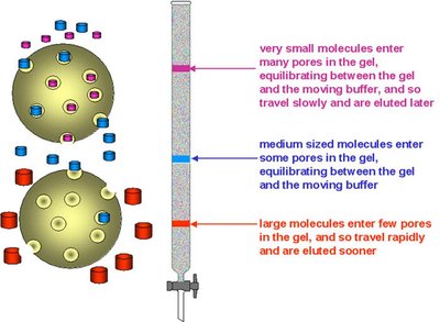
This method separates proteins by size using porous beads. Large proteins cannot enter the pores and elute first, while small proteins enter the pores and elute later.
Affinity Chromatography
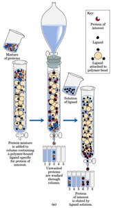
Affinity chromatography exploits specific interactions between a protein and a ligand attached to the matrix. Only proteins with affinity for the ligand are retained and later eluted with a solution containing free ligand.
Examples: Antigen-antibody, enzyme-substrate, receptor-ligand interactions.
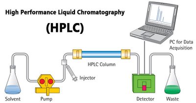
High-Performance Liquid Chromatography (HPLC)
HPLC uses high-pressure pumps to increase the speed and resolution of protein separation. It is highly effective but more expensive than traditional methods.
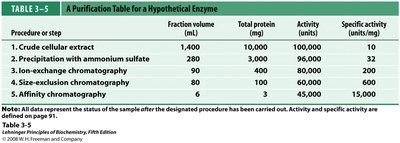
Quantification of Enzyme Purification
Enzyme purification is monitored by measuring activity and specific activity. Specific activity (units/mg protein) increases as purity increases and becomes constant when the enzyme is pure.
Procedure or Step | Fraction Volume (mL) | Total Protein (mg) | Activity (units) | Specific Activity (units/mg) |
|---|---|---|---|---|
Crude cellular extract | 1,400 | 10,000 | 100,000 | 10 |
Precipitation with ammonium sulfate | 280 | 3,000 | 96,000 | 32 |
Ion-exchange chromatography | 90 | 400 | 60,000 | 150 |
Size-exclusion chromatography | 80 | 100 | 48,000 | 480 |
Affinity chromatography | 6 | 3 | 45,000 | 15,000 |
Protein Electrophoresis and Analytical Techniques
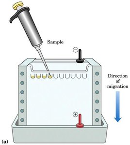
Electrophoresis
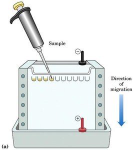
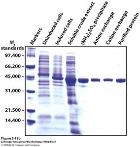
Electrophoresis separates proteins based on their charge and size in an electric field, typically using polyacrylamide gels. SDS-PAGE (sodium dodecyl sulfate-polyacrylamide gel electrophoresis) separates proteins primarily by mass, as SDS denatures proteins and gives them a uniform negative charge-to-mass ratio.
Estimating Molecular Weight by Electrophoresis
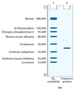
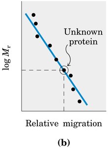
The molecular weight of an unknown protein can be estimated by comparing its migration to standards of known molecular weight and plotting log(Mr) versus relative migration.
Isoelectric Focusing (IEF)
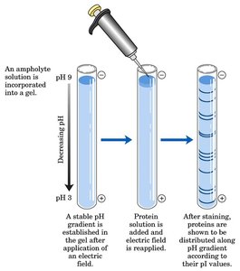
Isoelectric focusing separates proteins based on their isoelectric point (pI). Proteins migrate in a pH gradient until they reach the pH corresponding to their pI, where their net charge is zero.
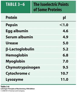
Protein | pI |
|---|---|
Pepsin | <1.0 |
Egg albumin | 4.6 |
Serum albumin | 4.9 |
Urease | 5.0 |
β-Lactoglobulin | 5.2 |
Hemoglobin | 6.8 |
Myoglobin | 7.0 |
Chymotrypsinogen | 9.5 |
Cytochrome c | 10.7 |
Lysozyme | 11.0 |
Two-Dimensional Electrophoresis
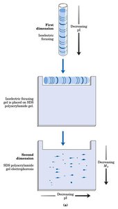
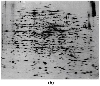
This technique combines isoelectric focusing and SDS-PAGE, allowing separation of proteins by both pI and molecular weight, thus resolving complex mixtures.
Protein Sequencing
Sanger Method
The Sanger method labels the amino-terminal residue with 1-fluoro-2,4-dinitrobenzene (FDNB), followed by hydrolysis to identify the labeled amino acid. It is limited to identifying the N-terminal residue only.
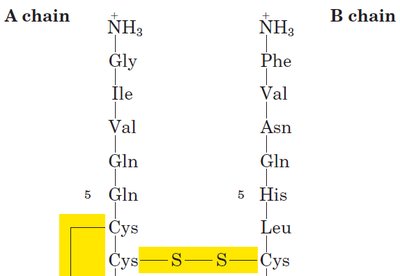
Edman Degradation
Edman degradation sequentially removes and identifies the N-terminal amino acid without hydrolyzing the entire peptide, allowing sequencing of up to 20–50 residues. Modern mass spectrometry can rapidly sequence short peptides.
Fragmentation and Disulfide Bond Analysis
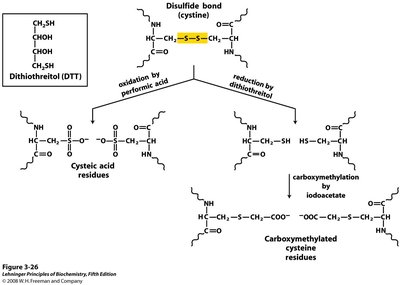
Large proteins are cleaved into smaller fragments using specific proteases or chemicals. Disulfide bonds must be reduced and alkylated to prevent reformation. Each fragment is sequenced, and their order is determined.
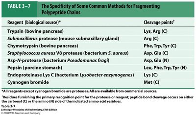
Reagent (source) | Cleavage Points |
|---|---|
Trypsin (bovine pancreas) | Lys, Arg (C) |
Submaxillaris protease (mouse submaxillary gland) | Arg (C) |
Chymotrypsin (bovine pancreas) | Phe, Trp, Tyr (C) |
Staphylococcus aureus V8 protease | Asp, Glu (C) |
Asp-N-protease (Pseudomonas fragii) | Asp, Glu (N) |
Pepsin (porcine stomach) | Leu, Phe, Trp, Tyr (N) |
Endoproteinase Lys C (Lysobacter enzymogenes) | Lys (C) |
Cyanogen bromide | Met (C) |
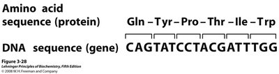
Protein Structure Overview
Levels of Protein Structure
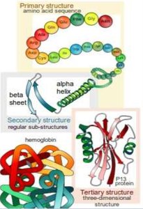
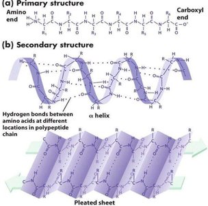
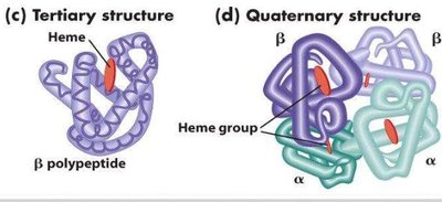
Proteins have four levels of structure: primary (amino acid sequence), secondary (α-helix and β-sheet), tertiary (three-dimensional folding), and quaternary (assembly of multiple polypeptides).