 뒤로
뒤로Protein Function: Oxygen Binding, Hemoglobin, Myoglobin, and Regulation
스터디 가이드 - 스마트 노트
 자료에 맞춘 맞춤형 노트, 핵심 정의, 예시, 맥락을 확장해 제공합니다.
Protein Function
Overview of Protein Function
Proteins serve a wide range of functions in biological systems, from structural support to catalysis and molecular transport. The function of a protein often depends on its ability to interact specifically and reversibly with other molecules, known as ligands. These interactions are central to processes such as oxygen transport and immune response.
Fibrous proteins provide structural support via stable quaternary interactions.
Globular proteins interact transiently with ligands, enabling dynamic physiological processes.
Ligand: Any molecule reversibly bound by a protein.
Binding site: The region on the protein complementary to the ligand in size, shape, charge, and hydrophobic/hydrophilic character.
Proteins are flexible, allowing conformational changes ("protein breathing") that facilitate ligand binding.
Induced Fit and Regulation
The binding of a ligand often induces a conformational change in the protein, enhancing the complementarity and affinity of the binding site. This induced fit mechanism is crucial for tight binding and specificity. In multisubunit proteins, conformational changes in one subunit can affect others, enabling regulation and cooperative binding.
Enzymes are proteins that bind and chemically transform substrates at their active site.
Regulation of protein-ligand interactions can occur via additional ligands that modulate protein conformation.
Oxygen Binding Proteins: Myoglobin and Hemoglobin
Heme Structure and Function
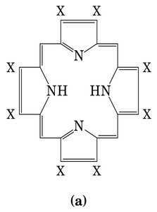
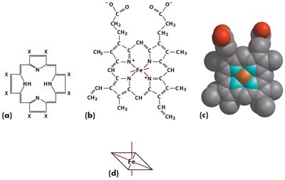
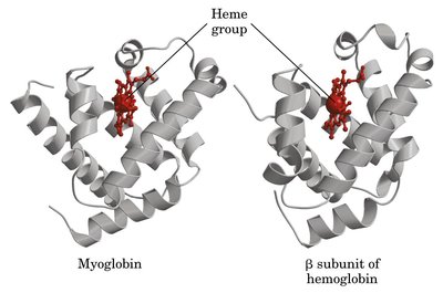
Oxygen transport and storage in vertebrates are mediated by myoglobin (muscle) and hemoglobin (blood), both of which utilize a prosthetic group called heme to bind oxygen. Heme is a complex organic ring structure (porphyrin) bound to a central iron ion (Fe2+).
Porphyrin consists of four pyrrole rings coordinated to Fe2+.
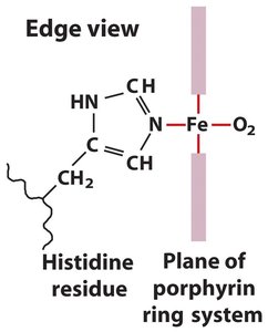
Iron in heme has six coordination bonds: four to nitrogen atoms in the porphyrin, one to a histidine residue (proximal His), and one to O2 (or other ligands).
Binding of O2 is reversible; binding of CO and NO is much stronger and toxic.
Myoglobin Structure and Function
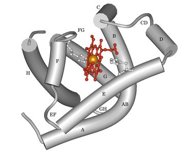
Myoglobin is a single polypeptide chain (153 amino acids) with a heme pocket formed mainly by E and F helices. It serves as an oxygen storage protein in muscle, binding O2 tightly and releasing it when needed.
Contains two histidine residues: proximal (anchors heme) and distal (facilitates O2 binding, reduces CO binding).
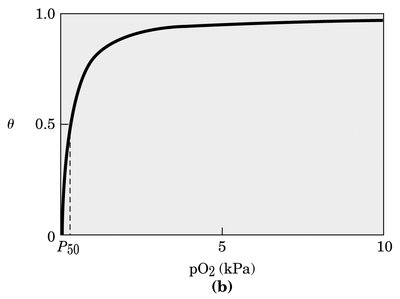
Myoglobin's oxygen binding curve is hyperbolic, indicating high affinity and insensitivity to small changes in O2 concentration.
Protein-Ligand Binding Equilibria
Binding Equilibrium and Constants
The reversible binding of a protein (P) to a ligand (L) is described by equilibrium constants:
Association constant:
Dissociation constant:
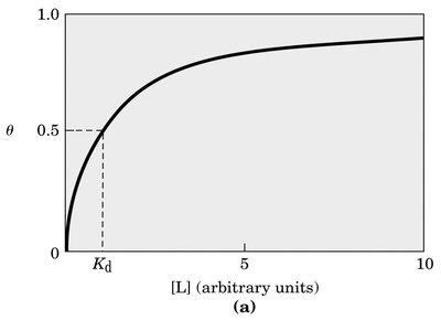
Fractional occupancy:
is the ligand concentration at which half of the binding sites are occupied.
Hemoglobin: Structure and Cooperative Binding
Hemoglobin Structure
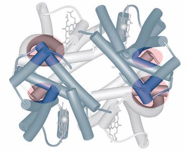
Hemoglobin is a tetramer composed of two alpha and two beta subunits, each with a heme group. Its quaternary structure allows for strong interactions between unlike subunits, enabling cooperative binding of oxygen.
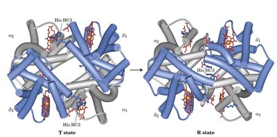
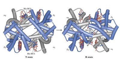
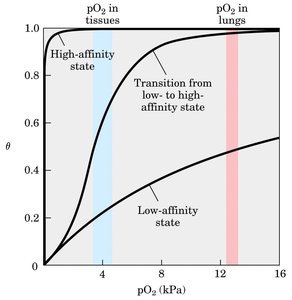
Exists in two states: T (tense, deoxy) and R (relaxed, oxy).
Oxygen binding induces a transition from T to R state, breaking ion pairs and narrowing the pocket between beta subunits.
Cooperative and Allosteric Binding
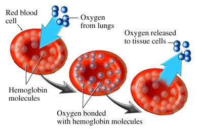
Hemoglobin displays cooperative binding, where binding of O2 to one subunit increases the affinity of the remaining subunits. This is reflected in its sigmoidal oxygen dissociation curve, making hemoglobin highly sensitive to changes in O2 concentration.
Allosteric proteins: Binding of a ligand at one site affects binding at another site.
Homotropic modulation: Normal ligand and modulator are identical (e.g., O2 in hemoglobin).
Heterotropic modulation: Modulator is a different molecule (e.g., 2,3-BPG, H+, CO2).
Regulation of Oxygen Binding
Bohr Effect: Influence of pH and CO2
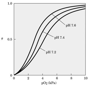
The Bohr effect describes how hemoglobin's affinity for oxygen decreases as pH drops (increased H+ concentration) and CO2 increases, facilitating oxygen release in tissues and uptake in lungs.
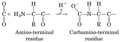
CO2 is converted to bicarbonate by carbonic anhydrase, increasing H+ and lowering pH.
Hemoglobin binds H+ and CO2 inversely to O2.
His HC3 of the beta subunit forms ion pairs stabilizing the T state when protonated.
2,3-Bisphosphoglycerate (BPG) Regulation
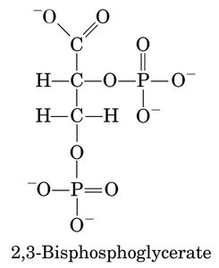
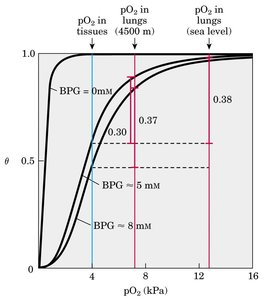
BPG is a heterotropic allosteric modulator that binds to hemoglobin and stabilizes the T state, reducing its affinity for oxygen. This adaptation is crucial for efficient oxygen delivery, especially at high altitudes.
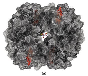
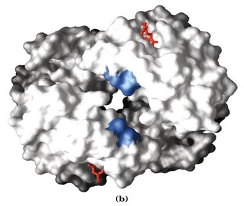
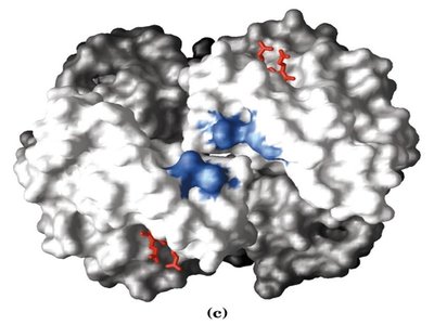
BPG binds in the pocket between beta subunits in the T state.
Increased BPG concentration lowers O2 affinity, enhancing oxygen release in tissues.
Fetal hemoglobin (α2γ2) has lower affinity for BPG, allowing greater O2 uptake from maternal blood.
Sickle Cell Anemia: Molecular Basis and Effects
Genetic Mutation and Hemoglobin Aggregation
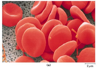
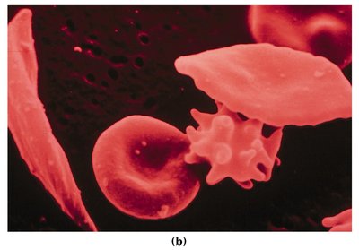
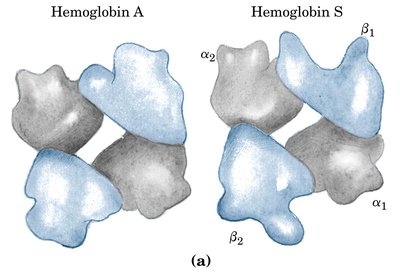
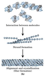
Sickle cell anemia is caused by a single amino acid substitution (Glu6 to Val6) in the beta chain of hemoglobin, creating a hydrophobic patch that promotes aggregation of deoxyhemoglobin S into insoluble fibers. This leads to abnormal, blade-shaped erythrocytes and serious medical complications.
Normal hemoglobin is soluble; hemoglobin S aggregates when deoxygenated.
Aggregation results in deformed erythrocytes, reduced oxygen transport, and increased risk of vascular blockage.
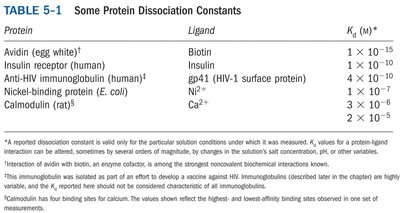
Summary Table: Protein Dissociation Constants
The following table summarizes dissociation constants (Kd) for various protein-ligand interactions, illustrating the range of affinities observed in biological systems.
Protein | Ligand | Kd (M) |
|---|---|---|
Avidin (egg white) | Biotin | 1 × 10-15 |
Insulin receptor (human) | Insulin | 1 × 10⁻¹⁰ |
Anti-HIV immunoglobulin | gp41 (HIV-1 surface protein) | 4 × 10⁻¹⁰ |
Nickel-binding protein (E. coli) | Ni2+ | 4 × 10⁻⁶ |
Calmodulin (rat) | Ca2+ | 2 × 10⁻⁵ |
