 뒤로
뒤로Protein Structure and Folding: Secondary, Tertiary, and Quaternary Organization
스터디 가이드 - 스마트 노트
 자료에 맞춘 맞춤형 노트, 핵심 정의, 예시, 맥락을 확장해 제공합니다.
Protein Structure and Folding
Overview of Protein Structure
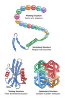
Proteins are essential biological macromolecules with complex structures that determine their diverse functions. Protein structure is organized hierarchically into four levels: primary, secondary, tertiary, and quaternary structure.
Primary structure: The linear sequence of amino acids in a polypeptide chain, linked by peptide bonds.
Secondary structure: Local folding patterns stabilized by hydrogen bonds, including α-helices and β-sheets.
Tertiary structure: The overall three-dimensional shape of a single polypeptide chain, formed by interactions among side chains.
Quaternary structure: The arrangement of multiple polypeptide subunits in a protein complex.
Covalent and Noncovalent Interactions in Protein Structure
Bond Strength and Stability
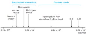
Covalent bonds, such as peptide bonds and disulfide bridges, are much stronger than noncovalent interactions (hydrogen bonds, ionic interactions, van der Waals forces, and hydrophobic effects). The stability of protein structure depends on the balance between these forces.
Covalent bonds: Provide strong, stable links (e.g., peptide bonds, disulfide bonds).
Noncovalent interactions: Include hydrogen bonds, ionic interactions, van der Waals forces, and hydrophobic effects; these are weaker but crucial for folding and flexibility.
Formation of the Peptide Bond
Peptide Bond Chemistry
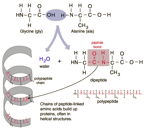
Amino acids are joined by peptide bonds through a condensation reaction, releasing water. The resulting polypeptide chain forms the backbone of proteins, which can adopt helical or extended conformations.
Peptide bond: Formed between the carboxyl group of one amino acid and the amino group of the next.
Polypeptide chain: Repeating backbone of N–Cα–C=O units, with variable side chains (R groups).
Secondary Structure: α-Helix
Discovery and Features of the α-Helix
The α-helix is a common protein secondary structure, first predicted by Pauling and colleagues using principles of chemistry and geometry. It is stabilized by regular hydrogen bonds between backbone atoms.
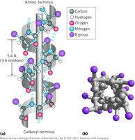
Helical backbone: The polypeptide backbone coils around a central axis, with R groups projecting outward.
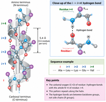
Hydrogen bonding: C=O of residue i forms a hydrogen bond with N–H of residue i+4.
Directionality: The α-helix has an N-terminus and a C-terminus.
Helix Geometry and Hydrogen Bonding Pattern
Each turn of the α-helix contains about 3.6 amino acids and is stabilized by a repeating pattern of hydrogen bonds. This arrangement allows for maximal hydrogen bonding without steric clashes.
Helix Handedness and Chirality
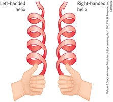
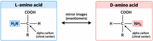
Most protein α-helices are right-handed due to the geometry of L-amino acids, which are the predominant form in biological systems. Left-handed helices are rare.
Factors Affecting α-Helix Stability
Charge: Like charges repel and can destabilize the helix; opposite charges can form stabilizing salt bridges.
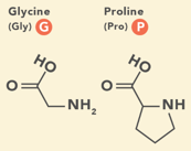
Size and shape: Bulky or rigid side chains (e.g., proline, glycine) can disrupt the helix.
Hydrophobicity: Hydrophobic side chains can stabilize helices by forming hydrophobic faces, especially in amphipathic helices.
Secondary Structure: β-Strands and β-Sheets
Structure and Organization of β-Strands
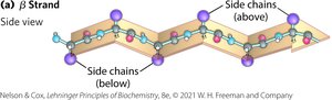
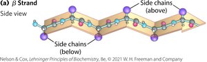
β-strands are extended polypeptide segments that align side-by-side to form β-sheets. The backbone is stretched in a zig-zag pattern, and side chains alternate above and below the plane of the sheet.
Amphipathic β-Sheets
Because side chains alternate, one face of a β-sheet can be hydrophobic while the other is polar or charged, allowing β-sheets to interact with different environments.
Parallel and Antiparallel β-Sheets
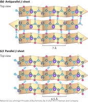
β-sheets can be formed from parallel or antiparallel β-strands. The orientation affects the hydrogen bonding pattern and the distance between strands.
Antiparallel β-sheet: Neighboring strands run in opposite directions; hydrogen bonds are more linear and stable.
Parallel β-sheet: Strands run in the same direction; hydrogen bonds are less linear.
β-Turns and Loops
Structure and Function of β-Turns
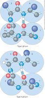
β-turns are short regions where the polypeptide chain reverses direction. They are stabilized by a hydrogen bond between the first and fourth residues. Proline and glycine are commonly found in β-turns due to their unique structural properties.
Dihedral Angles and the Ramachandran Plot
Backbone Dihedral Angles
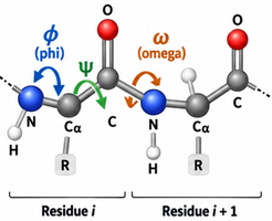
The conformation of the protein backbone is defined by three dihedral angles: φ (phi), ψ (psi), and ω (omega). These angles determine the possible secondary structures a region can adopt.
Ramachandran Plot
The Ramachandran plot shows the allowed combinations of φ and ψ angles for amino acid residues in proteins. Most residues fall within favored regions corresponding to α-helices and β-sheets, while other regions are disallowed due to steric clashes.
Key regions:
β-sheet region: φ ≈ –120° to –140°, ψ ≈ +120° to +140°
Right-handed α-helix region: φ ≈ –60°, ψ ≈ –45°
Left-handed α-helix region: φ ≈ +60°, ψ ≈ +40°
Folding Patterns: Motifs, Domains, and Folds
Protein Motifs and Folds
Secondary structures combine into motifs (e.g., β-α-β loops, β-barrels) and larger domains, which are the building blocks of tertiary structure. These recurring patterns help proteins achieve their functional three-dimensional shapes.
Tertiary and Quaternary Structure
Tertiary Structure
The tertiary structure is the overall three-dimensional arrangement of all atoms in a single polypeptide chain. It is stabilized by interactions among side chains, including hydrophobic interactions, hydrogen bonds, ionic interactions, and disulfide bonds.
Quaternary Structure
Quaternary structure refers to the assembly of multiple polypeptide chains (subunits) into a functional protein complex. Subunits can be identical or different and are held together by noncovalent interactions and sometimes covalent bonds.
Major Classes of Proteins Based on Structure
Fibrous Proteins
Long, strand-like or sheet-like; provide structural support (e.g., collagen, keratin).
Insoluble in water due to high hydrophobic residue content.
Globular Proteins
Compact, roughly spherical; perform diverse functions (e.g., enzymes, myoglobin, antibodies).
Folded into unique 3D shapes with hydrophobic cores and polar surfaces.
Membrane Proteins
Embedded in or associated with lipid membranes; contain hydrophobic regions that interact with the membrane interior.
Intrinsically Disordered Proteins
Lack a stable 3D structure under physiological conditions; enriched in flexible and charged residues.
Can adopt structure upon binding to partners, allowing regulatory flexibility.
Summary Table: Comparison of α-Helix and β-Sheet
Feature | α-Helix | β-Sheet |
|---|---|---|
Backbone hydrogen bonding | Within one strand (i → i+4) | Between neighboring strands |
Side chain orientation | Project outward from helix | Alternate above and below sheet |
Stabilizing interactions | Hydrogen bonds, salt bridges, hydrophobic effect | Hydrogen bonds, amphipathic faces |
Common destabilizers | Proline, glycine, bulky/charged side chains | Bulky/charged side chains, steric clashes |
Key Concepts
Protein structure is hierarchical: primary → secondary → tertiary → quaternary.
Secondary structures (α-helices, β-sheets) are stabilized mainly by backbone hydrogen bonds.
Side-chain properties (charge, size, hydrophobicity) influence folding and stability.
Backbone dihedral angles (φ, ψ) determine allowed conformations, visualized in Ramachandran plots.
Motifs and domains are modular units that build up functional proteins.
Protein structure determines function: fibrous proteins provide strength, globular proteins perform diverse functions, membrane proteins mediate transport/signaling, and intrinsically disordered regions allow flexible interactions.
