 뒤로
뒤로Protein Structure, Folding, and Function: Key Concepts in Biochemistry
스터디 가이드 - 스마트 노트
 자료에 맞춘 맞춤형 노트, 핵심 정의, 예시, 맥락을 확장해 제공합니다.
Protein Structure and Motifs
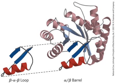
β-α-β Loop and α/β Barrel
Proteins are composed of recurring structural motifs that contribute to their overall three-dimensional architecture and function. Two important motifs are the β-α-β loop and the α/β barrel (also known as the TIM barrel). These motifs are commonly found in enzymes and play a critical role in stabilizing protein structure and facilitating catalytic activity.
β-α-β Loop: Consists of two parallel β-strands connected by an α-helix. This motif is a fundamental building block in many protein folds.
α/β Barrel: Formed by repeating β-α-β motifs, resulting in a barrel-like structure with eight parallel β-strands surrounded by eight α-helices. This structure is common in metabolic enzymes.
Example: Triose phosphate isomerase (TIM) is a classic example of a protein with an α/β barrel fold.
Intrinsically Disordered Proteins (IDPs)
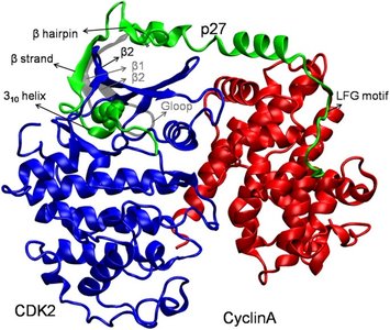
p27 and Protein-Protein Interactions
Some proteins or protein regions lack a fixed or ordered three-dimensional structure under physiological conditions. These are known as intrinsically disordered proteins (IDPs). IDPs often play regulatory roles and can interact with multiple partners, adopting structure upon binding.
p27: An IDP that regulates the cell cycle by binding to cyclin-dependent kinase 2 (CDK2) and CyclinA, inhibiting their activity.
Structural Adaptability: IDPs can transition from disorder to order upon binding, allowing for versatile regulatory functions.
Example: The interaction of p27 with CDK2 and CyclinA demonstrates how IDPs mediate complex cellular processes.
Protein Function: Hemoglobin and Oxygen Binding
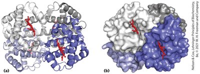
Binding of Oxygen by Heme
Hemoglobin is a classic example of a protein whose function is tightly linked to its structure. It binds oxygen via a heme prosthetic group, allowing for efficient oxygen transport in the blood.
Heme Group: An iron-containing porphyrin ring that reversibly binds oxygen molecules.
Cooperativity: Hemoglobin exhibits cooperative binding, where the binding of one oxygen molecule increases the affinity for subsequent oxygen molecules.
Example: The quaternary structure of hemoglobin enables allosteric regulation and efficient oxygen delivery to tissues.
Proteostasis: Protein Folding, Maintenance, and Degradation
Proteostasis Pathways
Proteostasis refers to the cellular processes that control the biogenesis, folding, trafficking, and degradation of proteins. Proper proteostasis is essential for cellular health and function.
Chaperones: Assist in the correct folding of nascent and misfolded proteins.
Degradation Pathways: Misfolded proteins are targeted for degradation via the ubiquitin-proteasome system or autophagy.
Aggregation: Failure in proteostasis can lead to protein aggregation and diseases such as Alzheimer's and Parkinson's.
Protein Denaturation and Stability
Denaturation of Proteins
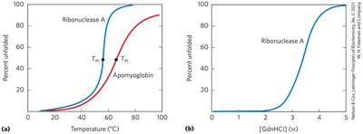
Denaturation is the process by which proteins lose their native structure due to external stress such as heat, pH changes, or chemical agents. This process is often reversible, but can sometimes lead to irreversible aggregation.
Thermal Denaturation: Increasing temperature disrupts non-covalent interactions, leading to protein unfolding.
Chemical Denaturation: Agents like guanidinium chloride (GdnHCl) or urea disrupt hydrogen bonds and hydrophobic interactions.
Melting Temperature (Tm): The temperature at which 50% of the protein is unfolded.
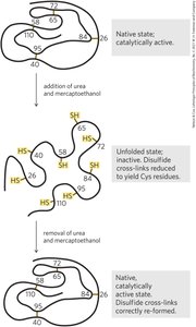
Reversible Denaturation of Ribonuclease A
Some proteins, such as ribonuclease A, can refold into their native, active conformation after denaturation if the primary structure remains intact. This demonstrates that all the information required for folding is contained in the amino acid sequence.
Disulfide Bonds: Reduction and reformation of disulfide bonds are critical for reversible folding.
Experiment: Removal of denaturants and reducing agents allows ribonuclease A to regain its native structure and catalytic activity.
Protein Folding Pathways
Schematics of Protein Folding
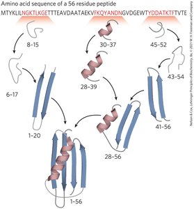
Protein folding is a highly ordered process that proceeds through a series of intermediate states. The sequence of amino acids determines the folding pathway and final structure.
Folding Intermediates: Local secondary structures form first, followed by the assembly of tertiary structure.
Folding Pathways: Multiple pathways may exist, but all lead to the native state with the lowest free energy.
Free Energy Funnel of Protein Folding
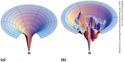
The folding of proteins can be visualized as a funnel-shaped energy landscape. The native state corresponds to the global minimum of free energy, while misfolded or partially folded states are higher in energy.
Energy Landscape: Smooth funnels indicate efficient folding, while rugged funnels represent kinetic traps and misfolded intermediates.
Thermodynamics: Folding is driven by the decrease in free energy as the protein adopts its native conformation.
Additional info: The concepts covered here are foundational for understanding protein structure-function relationships, folding diseases, and the design of therapeutic proteins.
