 뒤로
뒤로Protein Structure, Folding, Proteostasis, and Function
스터디 가이드 - 스마트 노트
 자료에 맞춘 맞춤형 노트, 핵심 정의, 예시, 맥락을 확장해 제공합니다.
Protein Structure and Proteostasis
Overview of Protein Structure
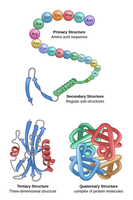
Proteins are essential macromolecules that perform a vast array of functions in biological systems. Their function is determined by their structure, which is organized into four hierarchical levels: primary, secondary, tertiary, and quaternary structure.
Primary Structure: The linear sequence of amino acids in a polypeptide chain, held together by peptide bonds.
Secondary Structure: Regular sub-structures such as α-helices and β-sheets, stabilized by hydrogen bonds.
Tertiary Structure: The overall three-dimensional folding of a single polypeptide chain, stabilized by various interactions including hydrophobic interactions, hydrogen bonds, ionic bonds, and disulfide bridges.
Quaternary Structure: The arrangement and interaction of multiple polypeptide subunits in a multi-subunit protein complex.
Proteostasis: Protein Homeostasis
Proteostasis refers to the cellular processes that control the biogenesis, folding, trafficking, and degradation of proteins. It is essential for maintaining a functional proteome and cellular health.
Protein Synthesis: Begins at the ribosome, producing a nascent polypeptide chain.
Folding: Newly synthesized proteins fold into their native, functional conformations, often with the assistance of molecular chaperones.
Modification and Trafficking: Proteins may undergo post-translational modifications and are transported to their correct cellular locations.
Degradation: Damaged or misfolded proteins are selectively degraded to prevent accumulation and cellular toxicity.
Protein Folding and Misfolding
Folding Pathways and Chaperones
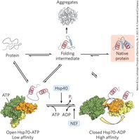
Protein folding is a complex process that can result in either the correct native structure or misfolded, non-functional forms. Chaperones are specialized proteins that assist in the folding process and prevent aggregation.
Folding Intermediates: Partially folded states that may require chaperone assistance to reach the native conformation.
Chaperone Function: Chaperones such as Hsp70 bind to unfolded or partially folded proteins, preventing aggregation and facilitating correct folding through ATP-dependent cycles.
Protein Misfolding and Cellular Consequences
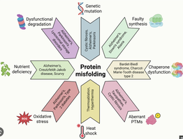
Proteins can misfold due to genetic mutations, environmental stress (e.g., heat shock), or chaperone dysfunction. Misfolded proteins may be refolded, degraded, or form toxic aggregates.
Refolding: Chaperones may attempt to remodel misfolded proteins.
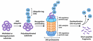
Degradation: The ubiquitin-proteasome system tags misfolded proteins for degradation into peptide fragments.
Aggregation: Persistent misfolded proteins can aggregate, forming oligomers or amyloid fibrils, which are associated with disease.
Anfinsen’s Experiment: Sequence Determines Structure
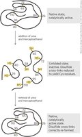
The classic experiment by Christian Anfinsen demonstrated that the information required for a protein to fold into its native structure is contained within its amino acid sequence. Denaturation and renaturation experiments with ribonuclease showed that removal of denaturants allows the protein to refold and regain activity, provided the primary sequence is intact.
Denaturation: Urea disrupts noncovalent interactions; β-mercaptoethanol reduces disulfide bonds.
Renaturation: Removal of denaturants allows correct refolding and reformation of disulfide bonds.
Protein Quality Control and Degradation Pathways
Ubiquitin-Proteasome System
The ubiquitin-proteasome system is the primary pathway for degrading small, soluble misfolded proteins. Proteins are tagged with ubiquitin, recognized by the 26S proteasome, and degraded into short peptides.
Ubiquitination: Covalent attachment of ubiquitin molecules to lysine residues on the substrate protein.
Proteasomal Degradation: The 19S regulatory particle recognizes ubiquitinated proteins, removes ubiquitin, and unfolds the substrate for degradation in the 20S core.
Autophagy
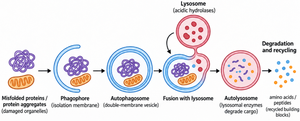
Autophagy is responsible for degrading larger protein aggregates, damaged organelles, and other cellular debris. The process involves the formation of a double-membrane autophagosome that engulfs the cargo and fuses with lysosomes for degradation.
Phagophore Formation: Initiates around the cargo to be degraded.
Autophagosome: A double-membrane vesicle that sequesters the cargo.
Fusion with Lysosome: Delivers the cargo to lysosomal enzymes for degradation into amino acids and peptides, which are recycled.
Protein Misfolding Diseases
Mechanisms of Protein-Folding Diseases
Protein misfolding can lead to a variety of diseases through several mechanisms:
Improper Degradation: Failure to degrade misfolded proteins leads to accumulation and toxicity.
Improper Localization: Misfolded proteins may be transported to incorrect cellular compartments.
Dominant-Negative Mutations: Mutant proteins interfere with the function of normal proteins.
Gain of Toxic Function: Misfolded proteins acquire new, harmful activities.
Amyloid Accumulation: Aggregation into amyloid fibrils is associated with neurodegenerative diseases such as Alzheimer's and Parkinson's.
Protein Function: Myoglobin and Hemoglobin
Oxygen-Binding Proteins (Globins)
Myoglobin and hemoglobin are globular proteins that bind oxygen via a heme prosthetic group. Myoglobin facilitates oxygen diffusion in muscle, while hemoglobin transports oxygen in the blood.
Myoglobin: Monomeric, high affinity for oxygen, stores and facilitates oxygen diffusion in muscle tissue.
Hemoglobin: Tetrameric, exhibits cooperative binding, efficiently loads oxygen in the lungs and releases it in tissues.
Heme Prosthetic Group
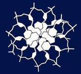
The heme group is a complex organic ring structure (protoporphyrin IX) with a central iron (Fe2+) atom that binds oxygen. The protein environment modulates the reactivity and binding properties of the heme.
Proximal Histidine (His93): Directly coordinates the iron atom.
Distal Histidine (His64): Stabilizes bound oxygen and prevents oxidation of Fe2+ to Fe3+.
Cooperative Oxygen Binding in Hemoglobin
Hemoglobin displays cooperative binding, meaning the binding of one oxygen molecule increases the affinity for subsequent oxygen molecules. This is reflected in a sigmoidal (S-shaped) oxygen binding curve.
T State (Tense): Low affinity for oxygen, stabilized by salt bridges and ion pairs.
R State (Relaxed): High affinity for oxygen, favored as oxygen binds and salt bridges are broken.
Allostery: Hemoglobin is an allosteric protein; ligand binding at one site affects binding at other sites.
Allosteric Modulation and the Bohr Effect
Hemoglobin's oxygen affinity is modulated by pH (Bohr effect), CO2, and other effectors. Lower pH (higher H+ concentration) decreases oxygen affinity, promoting oxygen release in metabolically active tissues.
Homotropic Modulator: Oxygen itself acts as a homotropic modulator, enhancing its own binding.
Heterotropic Modulators: H+ and CO2 decrease oxygen affinity, facilitating oxygen delivery to tissues.
Summary Table: Protein Folding and Quality Control Pathways
Process | Main Function | Key Players |
|---|---|---|
Chaperone-Assisted Folding | Assists correct folding, prevents aggregation | Hsp70, Hsp40, GroEL/GroES |
Ubiquitin-Proteasome System | Degrades misfolded/short-lived proteins | Ubiquitin, 26S proteasome |
Autophagy | Removes large aggregates, damaged organelles | Phagophore, autophagosome, lysosome |
