뒤로
뒤로A Tour of the Cell: Structure, Function, and Organization
스터디 가이드 - 스마트 노트
 자료에 맞춘 맞춤형 노트, 핵심 정의, 예시, 맥락을 확장해 제공합니다.
Chapter 6: A Tour of the Cell
Concept 6.1: Microscopy and Biochemistry in Cell Biology
Biologists use various microscopy and biochemical techniques to study cells, which are typically too small to be seen with the naked eye. These methods have enabled the discovery and detailed analysis of cellular structures and functions.
Microscopy:
Light Microscopy (LM): Uses visible light to illuminate specimens; suitable for living cells but limited in resolution.
Electron Microscopy (EM): Provides much higher resolution. Two main types:
Scanning Electron Microscopy (SEM): Produces detailed 3D images of cell surfaces.
Transmission Electron Microscopy (TEM): Reveals internal cell structures by passing electrons through thin sections.
Cell Fractionation: Involves breaking cells apart and separating organelles by centrifugation, allowing the study of individual cell components and their biochemical activities.
Key Terms: Magnification (enlargement of image) vs. Resolution (clarity of image).
Concept 6.2: Prokaryotic vs. Eukaryotic Cells; Animal vs. Plant Cells
Cells are classified as prokaryotic or eukaryotic based on their internal organization. Understanding these differences is fundamental to cell biology.
Prokaryotic Cells:
Domains: Bacteria and Archaea
DNA is located in the nucleoid (not membrane-bound).
Lack membrane-bound organelles.
Key structures: cell wall, plasma membrane, bacterial chromosome, nucleoid, ribosomes, flagella, fimbriae, capsule.
Eukaryotic Cells:
Domain: Eukarya (includes animals, plants, fungi, protists)
DNA is enclosed within a double-membrane nucleus.
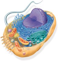
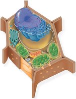
Contain membrane-bound organelles (e.g., mitochondria, ER, Golgi apparatus).
Animal vs. Plant Cells:
Plant cells have a cell wall, large central vacuole, and chloroplasts.
Animal cells have lysosomes, centrioles, and flagella (in some types).
Concept 6.3: The Nucleus, Chromosomes, and Ribosomes
The nucleus stores genetic information, while ribosomes are the sites of protein synthesis. Chromosomes are composed of chromatin (DNA + proteins).
Nucleus:
Enclosed by a double membrane (nuclear envelope) with nuclear pores for molecular transport.
Contains nucleolus (site of ribosome assembly) and chromatin.
Nuclear lamina and matrix provide structural support.
Ribosomes:
Composed of rRNA and proteins.
Types: Free ribosomes (cytosol; make proteins for cytoplasm) and Bound ribosomes (attached to ER/nuclear envelope; make proteins for membranes or export).
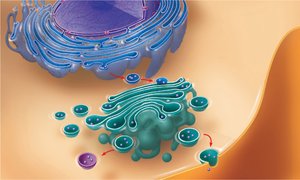
Concept 6.4: The Endomembrane System
The endomembrane system is a network of membranes involved in protein and lipid synthesis, modification, and transport.
Components: Nuclear envelope, endoplasmic reticulum (ER), Golgi apparatus, lysosomes, vacuoles, plasma membrane.
Endoplasmic Reticulum (ER):
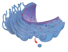
Rough ER: Studded with ribosomes; synthesizes proteins and glycoproteins.
Smooth ER: Lacks ribosomes; synthesizes lipids, metabolizes carbohydrates, detoxifies drugs/poisons.
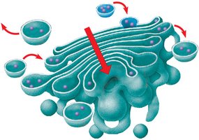
Golgi Apparatus: Modifies, sorts, and packages proteins and lipids for secretion or delivery to other organelles.
Lysosomes: Contain hydrolytic enzymes for intracellular digestion and recycling (autophagy).
Vacuoles:
Food vacuoles: Formed by phagocytosis.
Contractile vacuoles: Pump excess water out of cells (in protists).
Central vacuole (plants): Stores water, ions, and waste; maintains turgor pressure.
Concept 6.5: Mitochondria, Chloroplasts, and Peroxisomes
Mitochondria and chloroplasts are energy-transforming organelles, while peroxisomes carry out oxidation reactions.
Endosymbiont Theory: Mitochondria and chloroplasts originated as free-living prokaryotes engulfed by ancestral eukaryotic cells. Evidence includes double membranes, their own DNA, and ribosomes.
Mitochondria: Site of cellular respiration; converts glucose to ATP. Structure includes outer membrane, inner membrane (folded into cristae), intermembrane space, and matrix.
Chloroplasts: Site of photosynthesis in plants/algae; contains thylakoids (stacked into grana), stroma, and double membrane.
Peroxisomes: Break down fatty acids and detoxify harmful substances; produce hydrogen peroxide, then convert it to water.
Concept 6.6: The Cytoskeleton
The cytoskeleton is a dynamic network of protein fibers that provides structural support, facilitates cell movement, and organizes cellular components.
Types of Cytoskeletal Fibers:
Microtubules: Hollow tubes of tubulin; maintain cell shape, guide organelle movement, form cilia/flagella, separate chromosomes during cell division.
Microfilaments: Solid rods of actin; support cell shape, enable muscle contraction, and drive cell movement (e.g., amoeboid movement).
Intermediate Filaments: Fibrous proteins; provide mechanical support and anchor organelles.
Concept 6.7: Extracellular Components and Cell Junctions
Cells interact with their environment and each other through extracellular structures and specialized junctions.
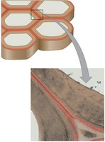
Plant Cells: Cell Wall Structure
Functions: Protects cell, maintains shape, prevents excessive water uptake.
Composition: Mainly cellulose, with hemicellulose and pectin.
Layers: Primary cell wall (thin/flexible), secondary cell wall (thicker, deposited after growth), middle lamella (pectin-rich layer between cells).
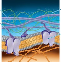
Animal Cells: Extracellular Matrix (ECM)
Components: Collagen fibers, proteoglycan complexes, fibronectin, integrins.
Functions: Provides structural support, regulates cell behavior, facilitates communication between cells and their environment.
Cell Junctions
Plant Cells: Plasmodesmata—channels that connect cytoplasm of adjacent cells, allowing passage of ions, molecules, and water.
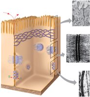
Animal Cells:
Tight Junctions: Seal cells together, preventing leakage of extracellular fluid.
Desmosomes: Anchor cells together into strong sheets.
Gap Junctions: Allow communication by permitting passage of small molecules and ions between cells.
Summary: Animal and Plant Cell Organization
Animal and plant cells share many organelles but also have unique structures that reflect their specialized functions.
Internal compartmentalization and extracellular structures enable cells to perform complex life processes efficiently.