 뒤로
뒤로A Tour of the Cell: Structure, Function, and Organization (8/16/26)
스터디 가이드 - 스마트 노트
 자료에 맞춘 맞춤형 노트, 핵심 정의, 예시, 맥락을 확장해 제공합니다.
Concept 6.1: Microscopy and Biochemistry in Cell Biology
Microscopy Techniques
Microscopy is essential for studying cells due to their small size. Two key properties of microscopes are magnification and resolution:
Magnification: The ratio of an object's image size to its real size. It determines how much larger the object appears. (think of the mirror's on your car beside you)
Resolution: The clarity of the image, or the minimum distance two points can be distinguished as separate. Higher resolution reveals more detail. (think of a camera)
Electron microscopy provides greater detail than light microscopy:
Scanning Electron Microscopy (SEM): Produces detailed 3D images of cell surfaces, showing depth and texture. (the surface level)
Transmission Electron Microscopy (TEM): Reveals internal structures by passing electrons through thin sections, but with less surface detail. ( the inside of the molecule)
Cell Fractionation
Cell fractionation separates cellular components by size and density using centrifugation. The smallest organelles isolated are ribosomes and microsomes.
Concept 6.2: Prokaryotic vs. Eukaryotic Cells; Animal vs. Plant Cells
Prokaryotic Cells
Prokaryotic cells (Domains: Archaea and Bacteria) lack a nucleus. Their DNA is found in the nucleoid, not enclosed by a membrane.
Cell wall: Rigid structure outside the plasma membrane
Plasma membrane: Encloses cytoplasm
Bacterial chromosome: DNA
Nucleoid: Region containing DNA
Ribosomes: Protein synthesis
Flagella: Locomotion
Surface Area-to-Volume Ratio
Cells are small to maximize surface area relative to volume, facilitating efficient exchange of materials. Microvilli increase surface area in intestinal cells.
As cell size increases, surface area-to-volume ratio decreases.
This limits the rate of material exchange, affecting cell function.
Concept 6.3: The Nucleus, Chromosomes, and Ribosomes
Nucleus Structure and Function
The nucleus is enclosed by a double-layered nuclear envelope with nuclear pores for molecular transport. The nuclear lamina and nuclear matrix provide structural support.
Chromatin: DNA and proteins; condenses into chromosomes during cell division. (crushes together)
Nucleolus: Site of ribosome assembly; visible when cells are not dividing. (public, when not dividing)
Ribosomes
Function: Protein synthesis (they check the proteins that are produced by the free ones)
Components: Large and small subunits
Free ribosomes: Located in cytosol; produce proteins for use within the cell.
Bound ribosomes: Attached to ER or nuclear envelope; produce proteins for secretion or membrane insertion. (they put in the proteins)
Concept 6.4: The Endomembrane System
Components and Functions
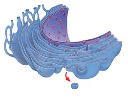
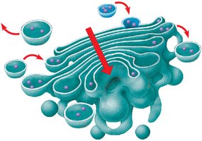
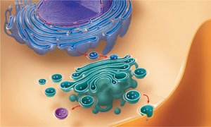
The endomembrane system includes the nuclear envelope, endoplasmic reticulum (ER), Golgi apparatus, lysosomes, vacuoles, and plasma membrane. It regulates protein traffic and metabolic functions.
Endoplasmic Reticulum (ER): Rough ER (with ribosomes) synthesizes proteins; Smooth ER (without ribosomes) synthesizes lipids, metabolizes carbohydrates, and detoxifies drugs.
Golgi Apparatus: Modifies, sorts, and packages proteins and lipids for secretion or delivery to other organelles. (like the Amazon for the membrane)
Lysosomes: Digestive organelles containing hydrolytic enzymes; acidic pH.
Vacuoles: Storage and transport; types include food, contractile, and central vacuoles (plants). (the U-Haul for food)
Proteins synthesized in the ER are transported in vesicles to the Golgi apparatus, where they are further processed and sorted.
Concept 6.5: Mitochondria and Chloroplasts
Endosymbiont Theory
The endosymbiont theory proposes that mitochondria and chloroplasts originated from prokaryotic cells that were engulfed by ancestral eukaryotes. Evidence includes:
Double membranes
Own DNA and ribosomes
Reproduction by binary fission (method of reproduction, ensuring genetic continuity)
Functions
Mitochondria: Site of cellular respiration; generates ATP. (ATP is like the gas for a truck the move the things around)
Chloroplasts: Site of photosynthesis; converts light energy to chemical energy. (remember this)
The highly folded inner membrane of mitochondria (cristae) and the stacked thylakoid membranes in chloroplasts increase surface area for energy conversion.
Concept 6.6: The Cytoskeleton
Types of Cytoskeletal Fibers
The cytoskeleton is a network of protein fibers that organizes cell structure and activities. Three main types:
Microtubules: Hollow tubes of tubulin; involved in cell shape, organelle movement, and cell division. (bones and fibers of a skeleton)
Microfilaments: Solid rods of actin; involved in cell movement and muscle contraction.(bones of a cytoskeleton)
Intermediate filaments: Fibrous proteins; provide structural support and maintain cell shape.
Concept 6.7: Extracellular Components and Cell Junctions
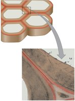
Plant Cell Walls
Plant cell walls provide structural support, protection, and regulate water intake. Composed mainly of cellulose.
Primary cell wall: Thin and flexible; secreted first. (weak)
Secondary cell wall: Deposited after primary wall; provides additional strength. (glue)
Middle lamella: Pectin-rich layer between cells; glues cells together. (Strong)
Animal Cell Extracellular Matrix (ECM)
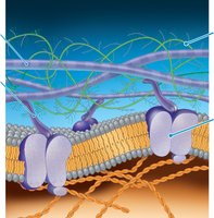
Animal cells lack cell walls but have an ECM composed of collagen, proteoglycans, fibronectin, and integrins. ECM provides structural support and mediates cell signaling.
Cell Junctions
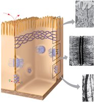
Cell junctions facilitate communication and adhesion between cells:
Tight junctions: Seal cells together, preventing leakage.(glue)
Desmosomes: Anchor cells together into strong sheets. (guys at the club door)
Gap junctions: Allow passage of ions and small molecules between cells. (people through the broader)
Cell Organization and Function
Animal and Plant Cell Structure
Animal and plant cells share many organelles but differ in some key structures:
Animal cells: Lack cell walls and chloroplasts; have centrioles and small vacuoles. (lack, small)
Plant cells: Have cell walls, chloroplasts, and a large central vacuole. (have, large)
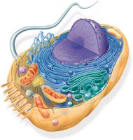
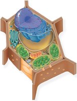
Internal organization allows eukaryotic cells to perform complex functions, including energy transformation, genetic information storage, and environmental interaction.
Additional info: The notes above expand on brief points with academic context, definitions, and examples, and include only images that are directly relevant to the explanation of each paragraph.
