 뒤로
뒤로Cell Communication and Signal Transduction Pathways (8/17/26)
스터디 가이드 - 스마트 노트
 자료에 맞춘 맞춤형 노트, 핵심 정의, 예시, 맥락을 확장해 제공합니다.
Q1. Label and describe the three stages of cell signaling using the example of epinephrine signaling in muscle cells.
Background
Topic: Cell Signaling Pathways
This question tests your understanding of the three main stages of cell signaling: reception, transduction, and response. These stages are fundamental to how cells detect and respond to external signals, such as hormones or neurotransmitters.

Key Terms and Concepts:
Signal Reception: The process by which a cell detects a signaling molecule from outside the cell.
Signal Transduction: The series of steps by which a signal on a cell’s surface is converted into a specific cellular response.
Cellular Response: The final action or change in cell activity triggered by the signal.
Step-by-Step Guidance
Identify the signaling molecule (in this case, epinephrine) and the target cell (muscle cell).
Describe how the signal is received: the epinephrine molecule binds to a specific receptor on the muscle cell membrane.
Explain what happens during signal transduction: the binding event activates relay molecules inside the cell, which transmit the signal through a cascade of molecular interactions.
Outline the cellular response: the signal ultimately activates an enzyme that breaks down glycogen, releasing glucose to fuel muscle activity.
Review the diagram and match each stage to its description, but stop before writing the final labels in the boxes.
Try solving on your own before revealing the answer!
Final Answer:
Signal reception: An epinephrine molecule binds to a receptor on a muscle cell.
Signal transduction: Relay molecules transmit the signal, ultimately activating an enzyme.
Cellular response: The enzyme breaks down glycogen, releasing glucose that fuels the leg muscles.
This sequence illustrates the universal stages of cell signaling: detection, relay, and response.
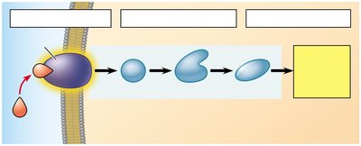
Q2. Label the missing parts in the signal transduction pathway diagram and explain what happens at each stage.
Background
Topic: Signal Transduction Pathways
This question focuses on the steps involved in a typical signal transduction pathway, where an external signal is relayed through a series of intracellular molecules, leading to a specific cellular response.
Key Terms and Concepts:
Receptor: A protein on the cell surface or inside the cell that binds the signaling molecule. (bring together)
Relay Molecules: Proteins and other molecules that pass the signal from the receptor to the target within the cell. (bow and arrow)
Cellular Response: The end result of the signaling pathway, such as enzyme activation or gene expression. (good, normal gene expression. bad, would be an extra chromosome)
Step-by-Step Guidance
Look at the first box: this is where the signaling molecule binds to the receptor on the cell membrane.
Follow the arrows: the signal is passed through a series of relay molecules inside the cell (signal transduction).
The final box represents the cellular response, which is the outcome of the pathway (e.g., activation of an enzyme or change in gene expression).
Think about what each box should be labeled based on the sequence: reception, transduction, and response.
Before filling in the labels, review the function of each stage and how the signal is relayed from the membrane to the cellular machinery.
Try solving on your own before revealing the answer!
Final Answer:
The boxes should be labeled (from left to right):
Signal Reception
Signal Transduction
Cellular Response
This diagram shows the flow of information from the cell surface to the inside, resulting in a specific cellular change.
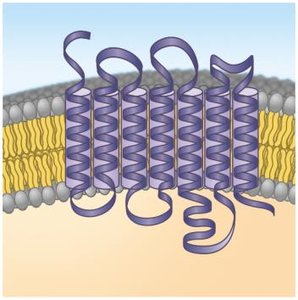
Q3. Label the structural features of a G-protein-coupled receptor (GPCR) shown in the diagram.
Background
Topic: Membrane Receptors – GPCRs
This question is about the structure of G-protein-coupled receptors, a major class of cell surface receptors involved in many signaling pathways.
Key Terms and Concepts:
GPCR: A receptor with seven transmembrane alpha helices that interacts with G proteins inside the cell.
Signaling Molecule Binding Site: The region where the ligand attaches to the receptor. (the cable that connects to a mic)
G Protein Interaction Site: The part of the receptor that interacts with the G protein on the cytoplasmic side. (the camera that the cable attaches to)
Step-by-Step Guidance
Identify the seven transmembrane alpha helices in the diagram; these are characteristic of GPCRs.
Locate the extracellular region where the signaling molecule would bind (usually at the top of the helices).
Find the intracellular region where the receptor interacts with the G protein (typically at the bottom of the helices).
Think about how the structure allows the receptor to transmit a signal from outside to inside the cell.
Prepare to label the binding site for the signaling molecule and the segment that interacts with the G protein, but stop before writing the final labels.
Try solving on your own before revealing the answer!
Final Answer:
Signaling molecule binding site: Located on the extracellular side of the receptor.
Seven transmembrane alpha helices: Span the membrane, forming the core structure of the GPCR.
G protein interaction site: Located on the intracellular side, where the receptor interacts with the G protein.
These features enable the GPCR to detect external signals and activate internal signaling pathways.
