 뒤로
뒤로Cell Division, Mitosis, and Meiosis: The Cellular Basis of Reproduction and Inheritance
스터디 가이드 - 스마트 노트
 자료에 맞춘 맞춤형 노트, 핵심 정의, 예시, 맥락을 확장해 제공합니다.
Cell Division: The Cellular Basis of Reproduction and Inheritance
Introduction to Cell Division
Cell division is a fundamental process by which a single parent cell divides to produce two or more daughter cells. This process is essential for reproduction, growth, and tissue repair in all living organisms.
Binary Fission: A type of cell division in prokaryotes (bacteria and archaea) where one cell splits into two identical cells.
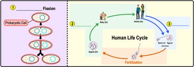
Mitosis: Eukaryotic cell division that produces genetically identical somatic (body) cells. Human somatic cells are diploid (2n), containing two copies of each chromosome.
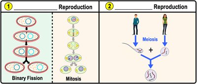
Meiosis: Eukaryotic cell division that produces haploid (n) gametes (sex cells). Human gametes have one copy of each chromosome.
Asexual vs. Sexual Reproduction
Organisms reproduce either asexually or sexually, each with distinct genetic outcomes.
Asexual Reproduction: Involves only one parent and produces genetically identical offspring. Examples include binary fission and mitosis.
Sexual Reproduction: Involves two parents, combining genetic material to produce genetically diverse offspring. Meiosis is the key process in sexual reproduction.
Importance of Cell Division
Cell division is crucial for:
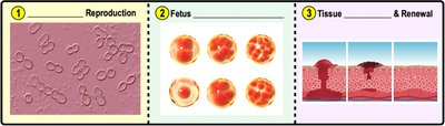
Reproduction: Making new organisms.
Fetal Development: Growth of multicellular organisms.
Tissue Repair: Renewal and healing of tissues.
Organization of DNA in the Cell
Genome and Chromatin Structure
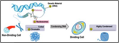
The genome is the complete set of a cell's DNA. DNA is organized with proteins called histones into nucleosomes, which further coil to form chromatin or chromosomes depending on the cell's state.
Chromatin: Loosely packed DNA in non-dividing cells.
Chromosomes: Highly condensed DNA in dividing cells.
DNA Replication and Chromosome Structure
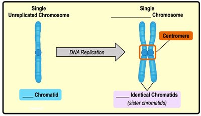
Before cell division, DNA is replicated to ensure each daughter cell receives a complete set of genetic material.
Chromatid: One half of a replicated chromosome, joined to its sister chromatid at the centromere.
Sister Chromatids: Genetically identical chromatids attached by a centromere.
The Cell Cycle
Phases of the Cell Cycle
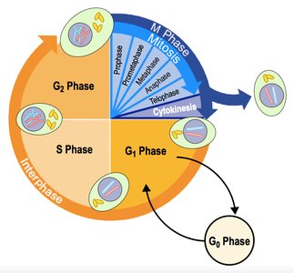
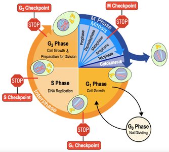
The cell cycle describes the sequence of events from cell formation to division. It consists of two major phases:
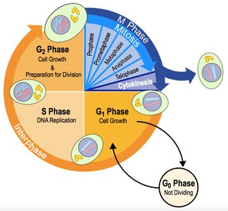
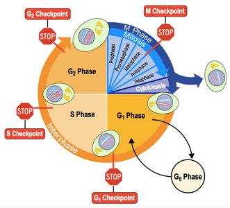
Interphase: Non-dividing phase for cell growth, DNA replication, and production of organelles and enzymes. Subdivided into G1 (growth), S (DNA synthesis), G2 (growth/preparation for division), and G0 (non-dividing).
M Phase (Mitotic Phase): Dividing phase including mitosis (nuclear division) and cytokinesis (cytoplasmic division).
Interphase Details
Interphase is the longest phase of the cell cycle, where the cell grows, replicates DNA, and prepares for division.
G1 Phase: Cell growth and normal function.
S Phase: DNA replication and centrosome duplication.
G2 Phase: Further growth and preparation for mitosis.
G0 Phase: Cells exit the cycle and do not divide.
Centrosomes and Mitotic Spindle
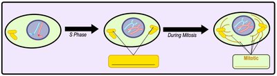
During S phase, centrosomes are replicated. They organize the mitotic spindle, which separates chromosomes during mitosis.
Centrosome: Microtubule-organizing center.
Mitotic Spindle: Microtubule structure that moves chromosomes.
Phases of Mitosis
Overview of Mitosis
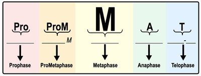
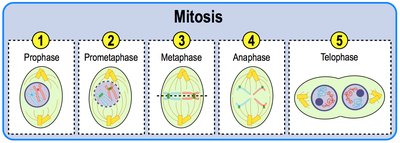
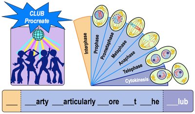
Mitosis is the process of dividing the nucleus and genetic material of a somatic cell, resulting in two genetically identical daughter cells. It consists of five phases:
Prophase
Prometaphase
Metaphase
Anaphase
Telophase
Prophase
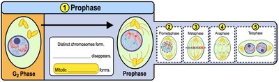
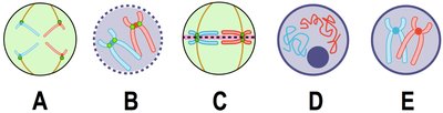
Chromatin condenses into visible chromosomes, nucleolus disappears, and centrosomes move to opposite poles, forming the mitotic spindle.
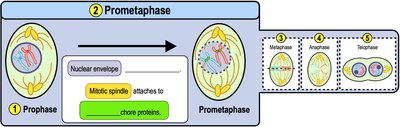
Prometaphase
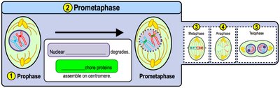
Nuclear envelope breaks down, exposing chromosomes. Spindle fibers attach to kinetochores at the centromere.
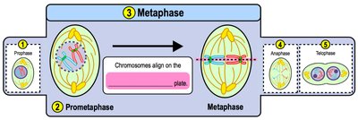
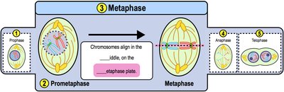
Metaphase
Chromosomes align at the cell's equator (metaphase plate), with spindle fibers attached to each chromatid.
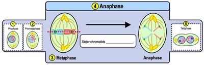
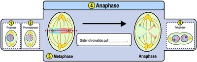
Anaphase
Sister chromatids are pulled apart toward opposite poles by shortening spindle fibers.
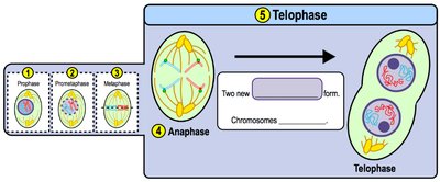
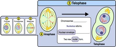
Telophase
Chromosomes decondense, spindle disassembles, and nuclear envelope reforms, creating two nuclei.
Cytokinesis
Animal Cell Cytokinesis
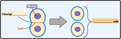
In animal cells, cytokinesis occurs via a cleavage furrow formed by actin microfilaments and myosin, dividing the cytoplasm.
Plant Cell Cytokinesis
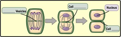
Plant cells form a cell plate from Golgi-derived vesicles, which develops into a new cell wall separating the daughter cells.
Cell Cycle Regulation
Checkpoints and Growth Factors
Cell division is regulated by growth factors and cell cycle checkpoints, which ensure proper progression and prevent errors.
Checkpoints: G1, S, G2, and M checkpoints monitor DNA integrity, replication, and chromosome alignment.
p53 Protein: Triggers repair or apoptosis if errors are detected.
Cancer: Results from cells ignoring checkpoints.
Genes, Alleles, and Chromosome Ploidy
Genes and Alleles
Genes are DNA segments encoding proteins for traits. Alleles are alternative versions of a gene, often represented by letters (e.g., B for blue eyes, b for brown eyes).
Haploid vs. Diploid Cells
Ploidy refers to the number of chromosome sets in a cell:
Haploid (n): One set of chromosomes (gametes).
Diploid (2n): Two sets of chromosomes (somatic cells).
Homologous Chromosomes and Karyotypes
Homologous Chromosomes
Homologous chromosomes are pairs similar in size and shape, carrying the same genes but possibly different alleles. Humans have 23 pairs: 22 autosomes and 1 pair of sex chromosomes (XX or XY).
Life Cycle of Sexual Reproducers
Meiosis and Fertilization
Sexual life cycles involve mitosis, meiosis, and fertilization. Meiosis produces haploid gametes, which fuse during fertilization to form a diploid zygote.
Meiosis: Process and Genetic Variation
Meiosis I and II
Meiosis consists of two rounds:
Meiosis I: Separates homologous chromosomes, reducing ploidy.
Meiosis II: Separates sister chromatids, similar to mitosis.
Genetic Variation in Meiosis
Meiosis creates genetic diversity through:
Crossing Over: Exchange of genetic material between homologous chromosomes during Prophase I.
Independent Assortment: Random alignment of chromosome pairs during Metaphase I. Number of combinations: (where n = haploid number of chromosomes).
Nondisjunction
Nondisjunction is an error where chromosomes fail to separate, resulting in aneuploid cells (extra or missing chromosomes), which can cause genetic disorders.
Mitosis vs. Meiosis Review
Comparison Table
Feature | Mitosis | Meiosis |
|---|---|---|
Cell Type | Somatic | Germ |
Number of Divisions | 1 | 2 |
Daughter Cells | 2, identical | 4, genetically diverse |
Ploidy | Diploid or Haploid | Haploid only |
Function | Growth, repair | Sexual reproduction |
Crossing Over | No | Yes |
Key Differences
Mitosis produces identical cells for growth and repair; meiosis produces genetically diverse gametes for reproduction.
Meiosis involves crossing over and independent assortment, increasing genetic variation.
Mitosis can occur in diploid or haploid cells; meiosis only in diploid cells.
Equational vs. Reductional Division
Equational Division: Daughter cells have the same chromosome number as the parent (mitosis, meiosis II).
Reductional Division: Daughter cells have half the chromosome number of the parent (meiosis I).
Additional info: These notes expand on brief points with academic context, definitions, and examples to provide a comprehensive study guide for General Biology students.
