 뒤로
뒤로Comprehensive Study Guidance for Cell Structure, Function, and Microscopy in General Biology
스터디 가이드 - 스마트 노트
 자료에 맞춘 맞춤형 노트, 핵심 정의, 예시, 맥락을 확장해 제공합니다.
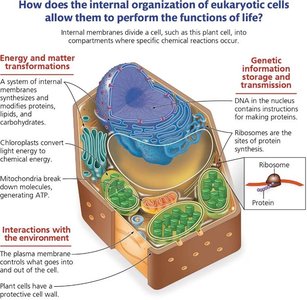
Q1. Describe an overview of key cell structures that are necessary to perform life functions.
Background
Topic: Cell Structure and Function
This question tests your understanding of the essential components found in all cells and their roles in maintaining life processes.
Key Terms:
DNA: Genetic material that directs cellular activities.
Ribosomes: Sites of protein synthesis.
Plasma Membrane: Regulates entry and exit of substances.
Cytoplasm: Internal fluid where organelles are suspended.
Step-by-Step Guidance
Identify the universal cell structures: DNA, ribosomes, plasma membrane, and cytoplasm.
Explain the function of each structure in supporting life processes (e.g., DNA for genetic information, ribosomes for protein synthesis).
Discuss why each structure is necessary for cell survival and function.
Consider how these structures interact to maintain cellular homeostasis.
Try solving on your own before revealing the answer!
Final Answer:
All cells contain DNA (for genetic information), ribosomes (for protein synthesis), a plasma membrane (for regulating entry/exit), and cytoplasm (for housing organelles and nutrients). These structures are essential for life because they enable cells to produce proteins, store and transmit genetic information, and interact with their environment.
Q2. Using the table, give the specific cell size ranges for the following and rank them smallest to largest: atom, ribosomes, small bacteria, mitochondria, animal cells, chicken egg.
Background
Topic: Cell Size and Scale
This question tests your ability to compare the sizes of various biological structures and understand their relative scale.
Key Terms:
Atom: Smallest unit of matter.
Ribosome: Organelle for protein synthesis.
Bacteria: Single-celled organisms.
Mitochondria: Organelle for energy production.
Animal Cell: Eukaryotic cell.
Chicken Egg: Large single cell.
Step-by-Step Guidance
Review the size ranges for each structure (atoms are measured in angstroms, ribosomes in nanometers, bacteria in micrometers, etc.).
Arrange the structures from smallest to largest based on their typical size.
Compare the scale differences between each structure.
Rank the structures in order, noting the magnitude of difference between each.
Try solving on your own before revealing the answer!
Final Answer:
Smallest to largest: atom < ribosome < small bacteria < mitochondria < animal cell < chicken egg. Atoms are the smallest, while chicken eggs are the largest single cells.
Q3. Describe the difference between the different microscopes listed below. Give examples of the structures that are visible by each type of microscope and the rationale for their use.
Background
Topic: Microscopy and Cell Visualization
This question tests your understanding of the types of microscopes used in biology and their applications.
Key Terms:
Light Microscope: Uses visible light to view cells.
Fluorescence Microscope: Uses fluorescent dyes to highlight structures.
Electron Microscope: Uses electron beams for high resolution.
Scanning Electron Microscope (SEM): Views surface details.
Transmission Electron Microscope (TEM): Views internal structures.
Step-by-Step Guidance
Define each type of microscope and its principle of operation.
List examples of cell structures visible with each microscope (e.g., light microscope for cells, electron microscope for organelles).
Explain why certain microscopes are chosen for specific structures (resolution, detail, live/dead cells).
Compare the advantages and limitations of each microscope type.
Try solving on your own before revealing the answer!
Final Answer:
Light microscopes are used for viewing whole cells and tissues; fluorescence microscopes highlight specific molecules; electron microscopes (SEM and TEM) provide high-resolution images of cell surfaces and internal structures. SEM is best for surface detail, TEM for internal detail.
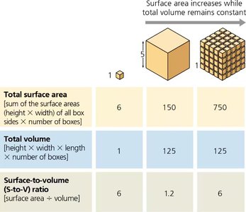
Q8. Why must cells be small to efficiently move materials back and forth across the membrane?
Background
Topic: Surface Area-to-Volume Ratio
This question tests your understanding of how cell size affects the efficiency of material transport and communication.
Key Terms and Formulas:
Surface Area: Total area of the cell membrane.
Volume: Space inside the cell.
Surface Area-to-Volume Ratio:
Step-by-Step Guidance
Recall that as a cell grows, its volume increases faster than its surface area.
Understand that a high surface area-to-volume ratio allows efficient exchange of materials.
Explain why larger cells have lower ratios and struggle to transport materials efficiently.
Discuss the consequences for cell function if the ratio becomes too low.
Try solving on your own before revealing the answer!
Final Answer:
Cells must be small because as size increases, the surface area-to-volume ratio decreases, making it harder for the cell to efficiently exchange materials with its environment. This is why cells divide when they reach a certain size.
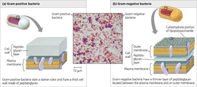
Q17. Using the figure, describe the structural differences in gram positive vs gram negative bacteria.
Background
Topic: Bacterial Cell Wall Structure
This question tests your ability to distinguish between gram-positive and gram-negative bacteria based on their cell wall composition.
Key Terms:
Gram-positive: Thick peptidoglycan layer.
Gram-negative: Thin peptidoglycan layer, outer membrane.
Peptidoglycan: Structural carbohydrate in bacterial cell walls.
Step-by-Step Guidance
Identify the main structural components of gram-positive and gram-negative bacteria.
Compare the thickness of the peptidoglycan layer in each type.
Note the presence or absence of an outer membrane in gram-negative bacteria.
Discuss how these differences affect antibiotic susceptibility.
Try solving on your own before revealing the answer!
Final Answer:
Gram-positive bacteria have a thick peptidoglycan layer and no outer membrane, while gram-negative bacteria have a thin peptidoglycan layer and an additional outer membrane. This structural difference affects how antibiotics like penicillin work.
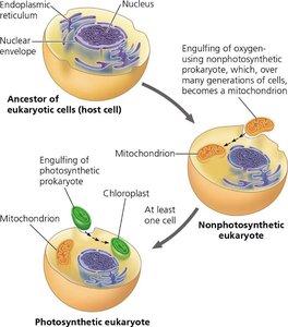
Q25. Describe the endosymbiosis theory and how the current eukaryotic cell may have derived with early origins with a prokaryotic cell.
Background
Topic: Evolution and Endosymbiosis Theory
This question tests your understanding of how eukaryotic cells evolved from prokaryotic ancestors through endosymbiosis.
Key Terms:
Endosymbiosis: One cell living inside another.
Mitochondria and Chloroplasts: Organelles thought to have originated from prokaryotes.
Step-by-Step Guidance
Explain the concept of endosymbiosis (engulfing of prokaryotes by ancestral eukaryotes).
Describe how mitochondria and chloroplasts are believed to have originated from prokaryotic cells.
Discuss the evidence supporting this theory (e.g., double membranes, own DNA).
Use the diagram to illustrate the process.
Try solving on your own before revealing the answer!
Final Answer:
The endosymbiosis theory proposes that eukaryotic cells originated when ancestral cells engulfed prokaryotes, which became mitochondria and chloroplasts. Evidence includes similarities in DNA, membranes, and reproduction between these organelles and prokaryotes.
Q50. What organelles or cell structures are likely malfunctioning in these diseases?
Background
Topic: Cell Structure and Disease
This question tests your ability to connect symptoms of diseases to malfunctioning organelles or cell structures.
Key Terms:
Cystic Fibrosis: Membrane transport proteins.
ALD: Peroxisomes (fatty acid breakdown).
Pompe: Lysosomes (macromolecule breakdown).
Kartagener: Cilia (movement of materials).
Step-by-Step Guidance
Read the symptoms and match them to the organelle's normal function.
Identify which organelle is affected based on the symptoms (e.g., mucus buildup suggests cilia malfunction).
Explain how the malfunction leads to the observed symptoms.
Fill in the table with the affected organelle for each disease.
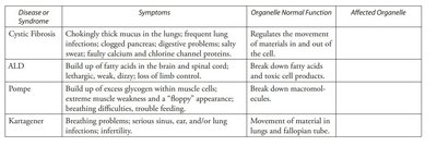
Try solving on your own before revealing the answer!
Final Answer:
Cystic Fibrosis: Plasma membrane transport proteins; ALD: Peroxisomes; Pompe: Lysosomes; Kartagener: Cilia. Each disease is linked to malfunction of a specific organelle responsible for the described symptoms.
