 뒤로
뒤로Comprehensive Study Guide for Cell Structure, Function, and Microscopy in General Biology
스터디 가이드 - 스마트 노트
 자료에 맞춘 맞춤형 노트, 핵심 정의, 예시, 맥락을 확장해 제공합니다.
Q1. Describe differences between light and electron microscopes, and how each is used to visualize the structure of cells.
Background
Topic: Cell Visualization Techniques
This question tests your understanding of the types of microscopes used in biology and their applications for viewing cellular structures.
Key Terms:
Light Microscope: Uses visible light to illuminate specimens; suitable for viewing living cells and larger organelles.
Electron Microscope: Uses beams of electrons for much higher resolution; suitable for viewing ultrastructure (very small details) of cells.
Fluorescence Microscope: Uses fluorescent dyes to highlight specific cell components.
Scanning Electron Microscope (SEM): Provides detailed surface images.
Transmission Electron Microscope (TEM): Reveals internal structures in great detail.
Step-by-Step Guidance
Identify the main differences in resolution and magnification between light and electron microscopes.
Consider which cell structures are visible with each type of microscope (e.g., nucleus, mitochondria, ribosomes).
Think about the advantages and limitations of each microscope (e.g., can you view living cells? What level of detail is possible?).
Review examples of cell structures that are best visualized with each microscope type.
Try solving on your own before revealing the answer!
Final Answer:
Light microscopes are used to view living cells and larger organelles (e.g., nucleus, mitochondria), but have limited resolution. Electron microscopes (TEM and SEM) provide much higher resolution, allowing visualization of smaller structures like ribosomes and cell membranes, but require dead, prepared samples. Fluorescence microscopes highlight specific molecules using dyes. SEM is best for surface details, TEM for internal structures.
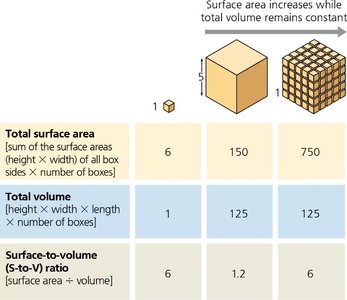
Q2. Explain how the surface area-to-volume ratio affects cell size and the efficiency of cellular processes, and why cells must remain small.
Background
Topic: Cell Size and Surface Area-to-Volume Ratio
This question tests your understanding of why cells are small and how their size impacts their function.
Key Terms and Formulas:
Surface Area: The total area covering the cell membrane.
Volume: The space inside the cell.
Surface Area-to-Volume Ratio:
Step-by-Step Guidance
Recall that as a cell grows, its volume increases faster than its surface area.
Calculate surface area and volume for a cube-shaped cell using and .
Compare how the ratio changes as the cell size increases.
Think about how this ratio affects the cell's ability to exchange materials with its environment.
Try solving on your own before revealing the answer!
Final Answer:
As cells increase in size, their surface area-to-volume ratio decreases, making it harder for the cell to efficiently exchange materials. This is why cells must remain small; a high ratio allows for efficient diffusion and transport across the membrane.
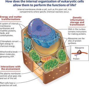
Q3. Describe the key structures and functions of key components of prokaryotic and eukaryotic cells.
Background
Topic: Cell Structure and Function
This question tests your knowledge of the basic features of prokaryotic and eukaryotic cells and their functions.
Key Terms:
Prokaryotic Cell: Lacks membrane-bound organelles; DNA in nucleoid; has cell wall, ribosomes, plasma membrane.
Eukaryotic Cell: Has membrane-bound organelles (nucleus, mitochondria, etc.); DNA in nucleus; more complex.
Ribosomes: Site of protein synthesis.
Plasma Membrane: Regulates entry/exit of materials.
Cytoplasm: Contains organelles and nutrients.
Step-by-Step Guidance
List the basic structures found in all cells (e.g., plasma membrane, ribosomes, cytoplasm, DNA).
Describe the function of each structure (e.g., ribosomes make proteins, plasma membrane controls entry/exit).
Compare prokaryotic and eukaryotic cells in terms of complexity and organelles.
Think about how these structures contribute to cell survival and function.
Try solving on your own before revealing the answer!
Final Answer:
All cells have a plasma membrane, ribosomes, cytoplasm, and DNA. Prokaryotes lack membrane-bound organelles and have DNA in a nucleoid, while eukaryotes have a nucleus and other organelles. Each structure plays a critical role in cell function and survival.
Q4. Describe how membrane-bound organelles help compartmentalize cellular functions.
Background
Topic: Cellular Compartmentalization
This question tests your understanding of how organelles allow cells to perform specialized functions efficiently.
Key Terms:
Membrane-bound organelles: Structures surrounded by membranes (e.g., nucleus, mitochondria, ER).
Compartmentalization: Separation of cellular processes into different areas.
Step-by-Step Guidance
Identify examples of membrane-bound organelles in eukaryotic cells.
Explain how each organelle's membrane creates a unique environment for specific reactions.
Consider how compartmentalization increases efficiency and prevents interference between processes.
Think about the consequences if organelles were not separated by membranes.
Try solving on your own before revealing the answer!
Final Answer:
Membrane-bound organelles compartmentalize cellular functions, allowing different metabolic processes to occur simultaneously and efficiently. This separation prevents interference and enables specialization within the cell.
Q5. Explain the structure and role of the cytoskeleton in supporting cell shape, enabling movement, and organizing cell structures.
Background
Topic: Cytoskeleton Structure and Function
This question tests your understanding of the cytoskeleton's components and their roles in cell function.
Key Terms:
Cytoskeleton: Network of protein filaments (microtubules, actin filaments, intermediate filaments).
Microtubules: Provide structural support, move organelles, and are components of cilia/flagella.
Actin Filaments: Enable cell movement and contraction.
Intermediate Filaments: Maintain cell shape and anchor organelles.
Step-by-Step Guidance
Identify the three main components of the cytoskeleton.
Describe the function of each component (e.g., microtubules move chromosomes, actin enables contraction).
Consider how the cytoskeleton supports cell shape and movement.
Think about what would happen if one or more components were missing.
Try solving on your own before revealing the answer!
Final Answer:
The cytoskeleton consists of microtubules, actin filaments, and intermediate filaments. It supports cell shape, enables movement, and organizes cell structures. Without these filaments, cells would lose shape, have impaired movement, and fail to transport materials properly.
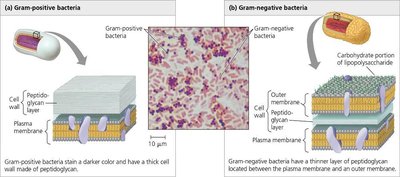
Q6. Using the figure, describe the structural differences in gram positive vs gram negative bacteria.
Background
Topic: Bacterial Cell Wall Structure
This question tests your ability to distinguish between gram-positive and gram-negative bacteria based on their cell wall structure.
Key Terms:
Gram-positive bacteria: Thick peptidoglycan layer, no outer membrane.
Gram-negative bacteria: Thin peptidoglycan layer, additional outer membrane.
Peptidoglycan: Structural carbohydrate in bacterial cell walls.
Step-by-Step Guidance
Examine the thickness of the peptidoglycan layer in each type of bacteria.
Identify the presence or absence of an outer membrane.
Consider how these differences affect staining and antibiotic susceptibility.
Think about why penicillin is more effective against gram-positive bacteria.
Try solving on your own before revealing the answer!
Final Answer:
Gram-positive bacteria have a thick peptidoglycan layer and no outer membrane, making them stain darker and more susceptible to antibiotics like penicillin. Gram-negative bacteria have a thin peptidoglycan layer and an outer membrane, which provides extra protection and makes them less susceptible to certain antibiotics.
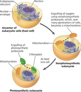
Q7. Describe the endosymbiosis theory and four pieces of evidence supporting it.
Background
Topic: Evolution of Eukaryotic Cells
This question tests your understanding of how mitochondria and chloroplasts originated from prokaryotic cells.
Key Terms:
Endosymbiosis Theory: Eukaryotic cells evolved by engulfing prokaryotic cells, which became mitochondria and chloroplasts.
Mitochondria and Chloroplasts: Organelles with similarities to prokaryotes.
Step-by-Step Guidance
Describe the process of a host cell engulfing prokaryotic cells.
List features shared by mitochondria/chloroplasts and prokaryotes (e.g., DNA, ribosomes).
Identify four pieces of evidence supporting the theory (e.g., double membranes, circular DNA, similar ribosomes, reproduction by binary fission).
Think about how these features are unique compared to other eukaryotic organelles.
Try solving on your own before revealing the answer!
Final Answer:
The endosymbiosis theory proposes that mitochondria and chloroplasts originated from prokaryotic cells engulfed by ancestral eukaryotes. Evidence includes: (1) both have double membranes, (2) both contain circular DNA, (3) both have prokaryote-like ribosomes, (4) both reproduce independently by binary fission.
Q8. Predict the malfunctioning organelle in the following cases: Low amounts of Ca2+ and large amounts of toxin and alcohol.
Background
Topic: Organelle Function and Disease
This question tests your ability to connect symptoms to organelle function.
Key Terms:
Smooth Endoplasmic Reticulum (SER): Detoxifies drugs/alcohol, stores calcium.
Step-by-Step Guidance
Identify which organelle is responsible for detoxification and calcium storage.
Connect the symptoms (low Ca2+, high toxins) to the function of the SER.
Consider what happens when the SER malfunctions.
Try solving on your own before revealing the answer!
Final Answer:
The malfunctioning organelle is the smooth endoplasmic reticulum (SER), which is responsible for detoxifying drugs/alcohol and storing calcium.
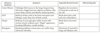
Q9. Using the chart, identify the affected organelle for each disease or syndrome listed.
Background
Topic: Cell Disease and Organelle Dysfunction
This question tests your ability to match symptoms and organelle function to specific diseases.
Key Terms:
Cystic Fibrosis: Plasma membrane
ALD: Peroxisome
Pompé: Lysosome
Kartagener: Cytoskeleton (microtubules)
Step-by-Step Guidance
Read the symptoms and normal function for each disease.
Match the symptoms to the organelle responsible (e.g., movement, breakdown of molecules).
Fill in the affected organelle for each disease.
Try solving on your own before revealing the answer!
Final Answer:
Cystic Fibrosis: Plasma membrane; ALD: Peroxisome; Pompe: Lysosome; Kartagener: Cytoskeleton (microtubules).
