 뒤로
뒤로Bio 100 LEC Chapter 16 Module 2
스터디 가이드 - 스마트 노트
 자료에 맞춘 맞춤형 노트, 핵심 정의, 예시, 맥락을 확장해 제공합니다.
Relationship Between Structure and Function in DNA
The double helix structure of DNA, as elucidated by Watson and Crick, is fundamental to its function as the genetic material. The specific base pairing (A with T, C with G) through hydrogen bonds allows for accurate copying of genetic information during cell division.
Base Pairing: Adenine (A) pairs with Thymine (T) via two hydrogen bonds; Cytosine (C) pairs with Guanine (G) via three hydrogen bonds.
Template Mechanism: Each strand of DNA can serve as a template for the synthesis of a new complementary strand, ensuring faithful transmission of genetic information.
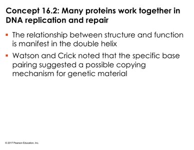
The Basic Principle: Base Pairing to a Template Strand
DNA replication relies on the separation of the two parental strands, each serving as a template for the synthesis of a new complementary strand. This process preserves the genetic code across generations.
Antiparallel Strands: DNA strands run in opposite directions (5' to 3' and 3' to 5').
Replication: Parental DNA separates, and new nucleotides are added according to base pairing rules, forming two identical DNA molecules.
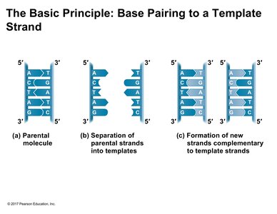
Models of DNA Replication
Conservative, Semiconservative, and Dispersive Models
Three models were proposed to explain how DNA replicates:
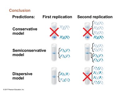
Conservative Model: The parental double helix remains intact, and an entirely new double helix is synthesized.
Semiconservative Model: Each daughter DNA molecule consists of one parental strand and one newly synthesized strand.
Dispersive Model: Each strand of both daughter molecules contains a mixture of old and new DNA.
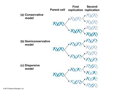
Experimental Evidence: The Meselson-Stahl Experiment
Matthew Meselson and Franklin Stahl designed an experiment using isotopes of nitrogen (N15 and N14) to distinguish between old and new DNA strands. Bacteria were grown in heavy (N15) media, then transferred to light (N14) media, and DNA was analyzed after each replication round using density gradient centrifugation.
Key Findings: After one replication, DNA showed intermediate density (hybrid), and after two replications, both hybrid and light DNA were present.
Conclusion: These results supported the semiconservative model of DNA replication.
Semiconservative Replication in Eukaryotes
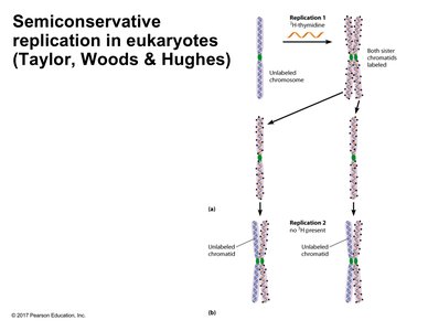
Further experiments in eukaryotes (e.g., broad bean root tips) using radioactive thymidine confirmed that DNA replication is semiconservative in both prokaryotes and eukaryotes. Autoradiography revealed that after one replication, both chromatids were labeled, but after two replications, only one chromatid per chromosome retained the label.
Mechanisms of DNA Replication
Origins of Replication: Prokaryotes vs. Eukaryotes
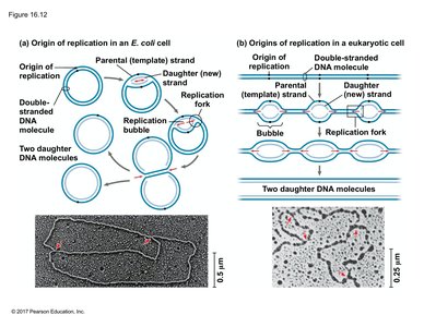
Replication begins at specific sites called origins of replication. Prokaryotes (e.g., E. coli) have a single origin, while eukaryotes have multiple origins due to their larger, linear chromosomes.
Replication Bubble: The region where DNA is unwound and replication occurs in both directions, forming replication forks.
Bidirectional Replication: Both prokaryotes and eukaryotes replicate DNA in two directions from each origin.
Proteins Involved in Initiating DNA Replication
Several proteins and enzymes are essential for the initiation and progression of DNA replication:
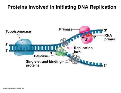
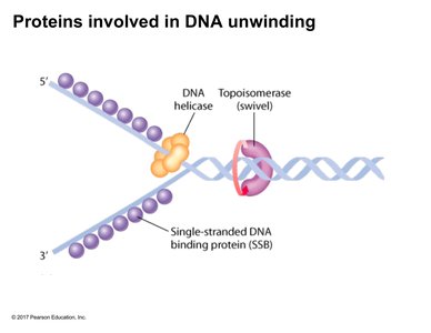
Helicase: Unwinds the DNA double helix using energy from ATP hydrolysis.
Single-Strand Binding Proteins (SSBs): Stabilize unwound DNA and prevent re-annealing or formation of hairpin structures.
Topoisomerase: Relieves supercoiling and torsional strain ahead of the replication fork by making transient cuts in the DNA.
Primase: Synthesizes short RNA primers needed to start DNA synthesis.
DNA Polymerase and the Synthesis of New DNA Strands
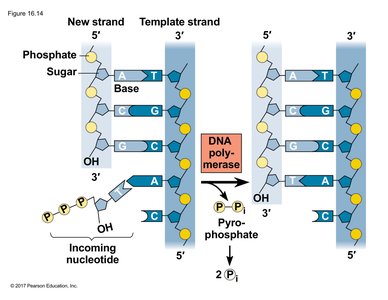
DNA polymerase catalyzes the addition of nucleotides to the growing DNA strand. It requires a primer with a free 3' hydroxyl group and adds nucleotides in the 5' to 3' direction, using deoxynucleoside triphosphates (dNTPs) as substrates. The energy for polymerization comes from the hydrolysis of the high-energy phosphate bonds in dNTPs.
Phosphodiester Bond Formation: Each new nucleotide is joined to the 3' end of the growing strand via a phosphodiester bond.
Pyrophosphate Release: Incorporation of a nucleotide releases pyrophosphate, which is hydrolyzed to drive the reaction forward.
Leading and Lagging Strand Synthesis
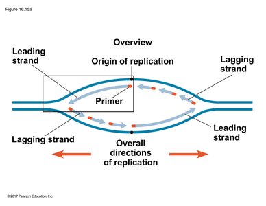
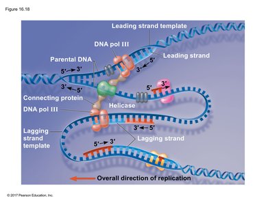
Because DNA polymerase can only synthesize in the 5' to 3' direction, replication is continuous on one strand (leading strand) and discontinuous on the other (lagging strand). The lagging strand is synthesized in short fragments called Okazaki fragments, each initiated by an RNA primer.
Leading Strand: Synthesized continuously toward the replication fork.
Lagging Strand: Synthesized discontinuously away from the replication fork in Okazaki fragments.
Proteins Involved in DNA Unwinding
Helicase, topoisomerase, and single-strand binding proteins work together to unwind and stabilize the DNA template during replication.
Leading Strand Synthesis: Stepwise Mechanism
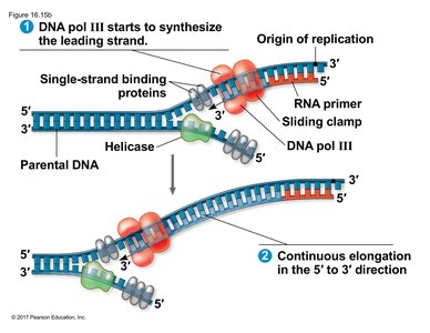
On the leading strand, primase synthesizes a single RNA primer, and DNA polymerase III extends the strand continuously. The sliding clamp protein holds DNA polymerase in place for efficient synthesis.
Lagging Strand Synthesis: Okazaki Fragments
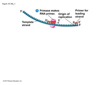
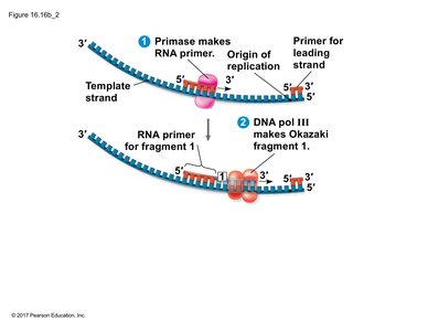
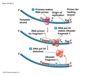
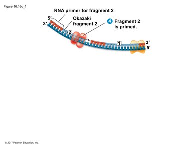
On the lagging strand, primase synthesizes multiple RNA primers. DNA polymerase III extends each fragment, which are later joined together. The process is discontinuous, with each fragment synthesized in the 5' to 3' direction.
Processing Okazaki Fragments
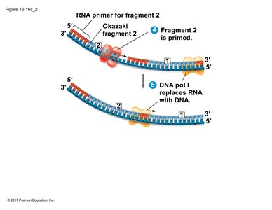
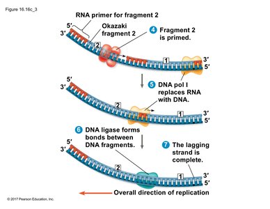
After DNA polymerase III synthesizes Okazaki fragments, DNA polymerase I removes the RNA primers and replaces them with DNA. DNA ligase then seals the nicks between fragments, forming a continuous DNA strand.
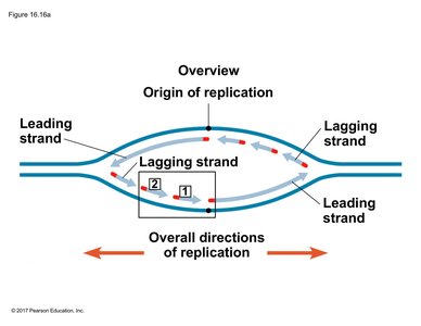
Replication Bubble Overview
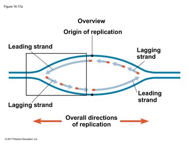
Within a replication bubble, leading and lagging strand synthesis occurs at each fork. The polarity of the DNA strands determines which regions are synthesized continuously or discontinuously.
The DNA Replication Machine (Replisome)
All proteins involved in DNA replication form a large complex called the replisome. This complex coordinates the synthesis of both leading and lagging strands, ensuring efficient and accurate DNA replication.
Proofreading and Repair of DNA
DNA Proofreading and Nucleotide Excision Repair
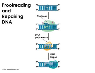
DNA polymerases have proofreading activity to correct errors during replication. Additional repair mechanisms, such as nucleotide excision repair (NER), remove and replace damaged DNA segments. Defects in repair pathways can lead to genetic disorders and increased cancer risk (e.g., xeroderma pigmentosum).
End Replication Problem and Telomeres
The End Replication Problem
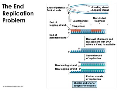
Linear chromosomes in eukaryotes face the end replication problem: after removal of the final RNA primer on the lagging strand, there is no way to fill in the resulting gap, leading to progressive shortening of chromosomes with each cell division.
Telomeres and Their Function
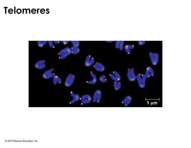
Telomeres are repetitive nucleotide sequences at the ends of linear chromosomes that protect genes from erosion. They do not prevent shortening but buffer the loss of essential genetic information. Telomere shortening is associated with cellular aging and senescence.
Telomerase and Chromosome Maintenance
In germ cells and certain stem cells, the enzyme telomerase extends telomeres, preventing their shortening and allowing cells to divide without limit. Telomerase is a reverse transcriptase that adds telomeric repeats to the 3' end of chromosomes, using an RNA template within the enzyme.
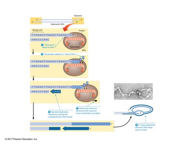
T-loop Structure: Telomeres can form protective T-loop structures at chromosome ends, further safeguarding genetic material.
