 뒤로
뒤로DNA Structure and Replication: Foundations of Molecular Biology
스터디 가이드 - 스마트 노트
 자료에 맞춘 맞춤형 노트, 핵심 정의, 예시, 맥락을 확장해 제공합니다.
DNA Structure & Replication
Introduction to DNA
Deoxyribonucleic acid (DNA) is the hereditary material in all living organisms, encoding the instructions necessary for life. While over 99% of human DNA is identical among individuals, small differences account for the diversity observed within populations. Understanding DNA structure and replication is essential for advances in medicine, genetics, and biotechnology.
Discovery of the Structure of DNA
Historical Context
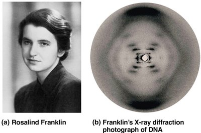
James Watson and Francis Crick proposed the double helix model of DNA in 1953, earning the Nobel Prize.
Rosalind Franklin contributed critical evidence through X-ray crystallography, revealing the helical structure of DNA. Her work was foundational but not fully credited during her lifetime.
DNA Structure
The Double Helix
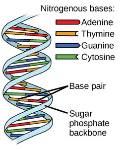
DNA consists of two antiparallel strands twisted into a double helix. The backbone is composed of alternating deoxyribose sugars and phosphate groups, while the interior contains nitrogenous base pairs.
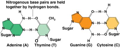
Base Pairing: Adenine (A) pairs with Thymine (T), and Guanine (G) pairs with Cytosine (C).
Antiparallel Orientation: One strand runs 5’ to 3’, the other 3’ to 5’.
Nucleotides: The Building Blocks of DNA
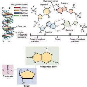
Each nucleotide is composed of a deoxyribose sugar, a phosphate group, and a nitrogenous base. The sequence of these bases encodes genetic information.
Hydrogen Bonds: Hold the two DNA strands together between complementary bases.
Directionality: DNA synthesis and reading always occur in the 5’ to 3’ direction.
Nitrogenous Bases: Purines and Pyrimidines
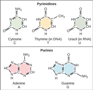
Nitrogenous bases are classified into two groups:
Pyrimidines: Single-ring structures (Cytosine, Thymine, and Uracil in RNA).
Purines: Double-ring structures (Adenine and Guanine).
Base Pairing Rule: Pyrimidines always pair with purines (A with T/U, G with C).
Base Pairing Specificity
The specificity of base pairing is due to the number of hydrogen bonds formed:
Adenine–Thymine: Two hydrogen bonds.
Guanine–Cytosine: Three hydrogen bonds (stronger pairing).
In RNA, Uracil replaces Thymine and pairs with Adenine.
DNA Replication
Semi-Conservative Model
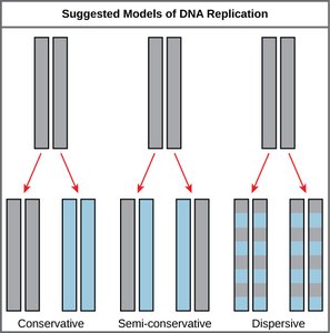
DNA replication follows the semi-conservative model, where each new DNA molecule consists of one parental and one newly synthesized strand. This was confirmed using isotopic labeling experiments.
Conservative Model: Parental DNA remains intact; new molecule is entirely new.
Semi-Conservative Model: Each daughter DNA has one old and one new strand.
Dispersive Model: Each strand is a mix of old and new DNA segments.
Major Steps and Key Enzymes in DNA Replication
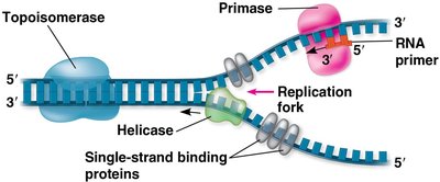
DNA replication is a highly coordinated process involving several enzymes and proteins:
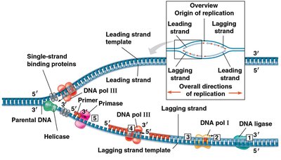
Origin of Replication: Specific sequence where replication begins.
Helicase: Unwinds the DNA double helix, creating a replication fork.
Single-Strand Binding Proteins: Stabilize unwound DNA strands.
Topoisomerase: Relieves tension ahead of the replication fork.
Primase: Synthesizes short RNA primers to provide a starting point for DNA synthesis.
Leading and Lagging Strands
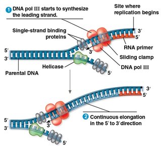
DNA polymerase III synthesizes new DNA in the 5’ to 3’ direction. Because the two template strands are antiparallel, replication proceeds differently on each:
Leading Strand: Synthesized continuously toward the replication fork.
Lagging Strand: Synthesized discontinuously away from the fork in short segments called Okazaki fragments, each requiring a new primer.
Okazaki Fragments and DNA Ligation
On the lagging strand, DNA polymerase I replaces RNA primers with DNA, and DNA ligase joins the Okazaki fragments to form a continuous strand.
Okazaki Fragments: Short DNA segments synthesized on the lagging strand.
DNA Ligase: Seals nicks between fragments, completing the strand.
Proofreading and DNA Repair
Nucleotide Excision Repair
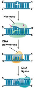
DNA replication is highly accurate, but errors can occur. Cells possess repair mechanisms to correct mistakes:
Nuclease: Removes the damaged or incorrect DNA segment.
DNA Polymerase: Fills in the correct nucleotides.
DNA Ligase: Seals the repaired section into the DNA strand.
Unrepaired mistakes become permanent mutations.
Telomeres and Replication of Chromosome Ends
Telomere Function and Telomerase
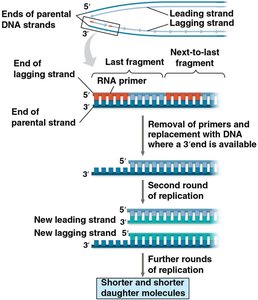
Each round of DNA replication shortens the ends of linear chromosomes. Telomeres, repetitive non-coding sequences at chromosome ends, protect genetic information from loss. In certain cells (germ cells, stem cells, cancer cells), the enzyme telomerase extends telomeres, counteracting shortening and maintaining chromosome integrity.
Telomeres: Buffer regions that prevent loss of essential genes.
Telomerase: Enzyme that adds telomeric repeats using an RNA template.
Telomere shortening is associated with cellular aging.
Summary Table: Key Enzymes and Functions in DNA Replication
Enzyme/Protein | Function |
|---|---|
Helicase | Unwinds DNA double helix |
Single-Strand Binding Proteins | Stabilize unwound DNA |
Topoisomerase | Relieves supercoiling ahead of fork |
Primase | Synthesizes RNA primers |
DNA Polymerase III | Adds nucleotides to new DNA strand |
DNA Polymerase I | Replaces RNA primers with DNA |
DNA Ligase | Joins Okazaki fragments |
Nuclease | Removes damaged DNA |
Telomerase | Extends telomeres in specific cells |
