뒤로
뒤로Bio 100 LEC Chapter 16 Module 1
스터디 가이드 - 스마트 노트
 자료에 맞춘 맞춤형 노트, 핵심 정의, 예시, 맥락을 확장해 제공합니다.
Chapter 16: The Molecular Basis of Inheritance
Historical Context and Scientific Inquiry
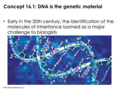
Early in the 20th century, the identification of the molecules responsible for inheritance was a major challenge for biologists. The discovery that DNA is the genetic material was not immediately obvious, as several types of large biological molecules were considered possible candidates. Through a series of experiments, scientists established DNA as the carrier of heritable information.
Experimental Evidence for DNA as Genetic Material
The Griffith Experiment: Bacterial Transformation
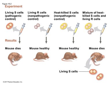
Frederick Griffith's experiment demonstrated the phenomenon of transformation in bacteria. He worked with two strains of Streptococcus pneumoniae: the pathogenic S (smooth) strain, which has a polysaccharide coat and causes pneumonia, and the non-pathogenic R (rough) strain, which lacks the coat and does not cause disease. Griffith found that when heat-killed S cells were mixed with living R cells and injected into mice, the mice died and living S cells were recovered, indicating that some "transforming principle" from the dead S cells converted R cells into pathogenic S cells.
S strain (smooth): Has a protective polysaccharide coat; causes disease.
R strain (rough): Lacks the coat; does not cause disease.
Heat-killed S strain: Non-viable; does not cause disease.
Mixture of heat-killed S and living R: Causes disease; living S cells recovered.
Conclusion: Heritable information can be transferred between bacteria.
Avery, MacLeod, and McCarty: Identifying the Transforming Principle
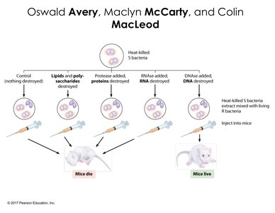
Building on Griffith's work, Avery, MacLeod, and McCarty sought to identify the specific molecule responsible for transformation. They treated cell-free extracts of heat-killed S bacteria with enzymes that destroyed proteins, RNA, or DNA. Only when DNA was destroyed did transformation fail to occur, indicating that DNA is the genetic material responsible for transformation.
Protease treatment: Proteins destroyed; transformation still occurs.
RNase treatment: RNA destroyed; transformation still occurs.
DNase treatment: DNA destroyed; transformation does not occur.
Conclusion: DNA is the molecule of inheritance in bacteria.
Evidence from Viral DNA: Bacteriophage Experiments
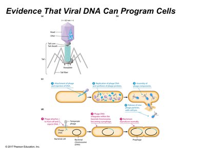
Bacteriophages (viruses that infect bacteria) provided further evidence for DNA as the genetic material. The viral life cycle involves the injection of genetic material into the host cell, which then directs the synthesis of new viral particles. The debate between DNA and protein as the genetic material was addressed using these viruses, which contain only DNA and protein.
Lytic cycle: Viral DNA is injected, directs synthesis of new phages, and host cell bursts.
Lysogenic cycle: Viral DNA integrates into host genome and is replicated with each cell division.
Conclusion: Viral DNA, not protein, programs the host cell.
The Hershey-Chase Experiment
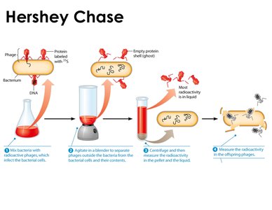
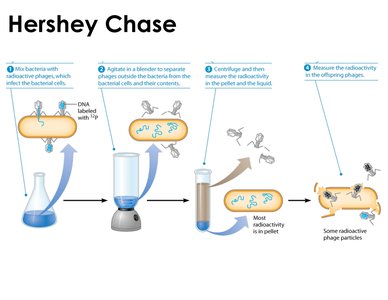
Alfred Hershey and Martha Chase used radioactive labeling to distinguish between DNA and protein in bacteriophages. In one experiment, phages were labeled with radioactive sulfur (found in proteins, not DNA); in another, with radioactive phosphorus (found in DNA, not proteins). After infection and separation, only radioactive phosphorus was found inside the bacteria and in new phages, confirming that DNA is the genetic material.
Radioactive sulfur (protein): Found in supernatant, not in bacterial cells.
Radioactive phosphorus (DNA): Found in pellet (bacterial cells) and in new phages.
Conclusion: DNA, not protein, is injected and directs viral replication.
DNA Structure and Composition
Chargaff’s Rules: Base Composition of DNA
Erwin Chargaff analyzed the base composition of DNA from various organisms and found two key patterns: (1) the amount of adenine (A) equals thymine (T), and the amount of guanine (G) equals cytosine (C); (2) the relative proportions of AT and GC pairs vary between species. These findings provided quantitative support for the double helix model.
A = T and G = C in any species.
AT/GC ratio: Varies between species, reflecting genetic diversity.
Implication: Base pairing is fundamental to DNA structure.
Building the Structural Model of DNA: X-ray Crystallography
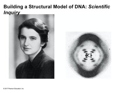
Rosalind Franklin's X-ray diffraction images revealed the repeating helical structure of DNA. Her work was pivotal in deducing the double helix model, showing that DNA has a regular, repeating pattern and that nucleotides can hydrogen bond under physiological conditions.
X-ray diffraction: Reveals helical structure and repeating patterns.
Hydrogen bonding: Enables base pairing between nucleotides.
Scientific impact: Franklin's work was foundational for Watson and Crick's model.
The Watson-Crick Model: Double Helix Structure
Watson and Crick synthesized previous findings to propose the double helix model of DNA. The structure consists of two antiparallel strands held together by hydrogen bonds between complementary bases (A-T and G-C). The helix has a diameter of 2 nm, with structural repeats every 0.34 nm and every 3.4 nm (one full turn every 10 base pairs). The sequence of one strand determines the sequence of the other, and the strands run in opposite directions (antiparallel).
Double helix: Two strands, antiparallel, held by hydrogen bonds.
Base pairing: A-T (2 hydrogen bonds), G-C (3 hydrogen bonds).
Structural repeats: 0.34 nm between bases, 3.4 nm per turn.
Complementarity: Sequence of one strand determines the other.
Antiparallel: One strand runs 5' to 3', the other 3' to 5'.
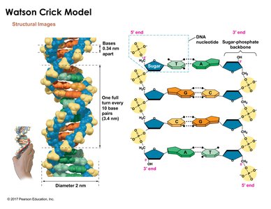
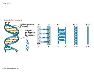
Simplified Representations of DNA Structure
DNA can be illustrated at various levels of detail, from the full helical structure to simplified two-dimensional diagrams. These representations highlight the sugar-phosphate backbone, nitrogenous base pairing, and the antiparallel orientation of the strands. In diagrams, terminal ends may fade to indicate that more DNA exists beyond the depicted region.
Helical model: Shows turns and backbone.
Base pairing diagrams: Focus on complementary bases.
Strand orientation: 5' and 3' ends marked; antiparallel arrangement.
Diagram conventions: Faded ends imply additional DNA not shown.