 뒤로
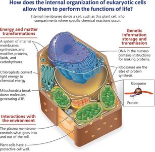
뒤로General Biology: Cell Structure, Function, and Microscopy Study Guide
스터디 가이드 - 스마트 노트
 자료에 맞춘 맞춤형 노트, 핵심 정의, 예시, 맥락을 확장해 제공합니다.
Q1. Describe an overview of key cell structures that are necessary to perform life functions.
Background
Topic: Cell Structure and Function
This question tests your understanding of the essential components found in all cells and their roles in maintaining life processes.
Key Terms:
Plasma Membrane: Boundary that regulates what enters and exits the cell.
Cytoplasm: Gel-like substance where cellular processes occur.
DNA: Genetic material that directs cell activities.
Ribosomes: Structures that synthesize proteins.
Step-by-Step Guidance
List the four basic structures found in all cells: plasma membrane, cytoplasm, DNA, and ribosomes.
Briefly describe the function of each structure (e.g., plasma membrane controls entry/exit, DNA stores genetic info, etc.).
Think about why each structure is essential for life (e.g., why is protein synthesis necessary?).
Consider how these structures interact to support cell survival and function.
Try solving on your own before revealing the answer!
Final Answer:
All cells have a plasma membrane (regulates entry/exit), cytoplasm (site of metabolic reactions), DNA (genetic instructions), and ribosomes (protein synthesis). These structures are essential for maintaining homeostasis, heredity, and cellular function.
Q2. Using the table to the right, give the specific cell size ranges for the following and rank them smallest to largest: Atom, Ribosomes, Small Bacteria, Mitochondria, Animal Cells, Chicken Egg.
Background
Topic: Cell Size and Microscopy
This question tests your ability to compare the sizes of biological structures and understand their scale.
Key Terms:
Micrometer (μm): Unit of length commonly used to measure cells.
Nanometer (nm): Unit of length used for very small structures like ribosomes.
Step-by-Step Guidance
Refer to the size ranges typically given for each structure (e.g., atoms are less than 1 nm, ribosomes ~20-30 nm, etc.).
Convert all sizes to the same unit (e.g., nm or μm) for easy comparison.
Arrange the structures in order from smallest to largest based on their size.
Double-check the order and size ranges using your textbook or class notes.
Try solving on your own before revealing the answer!
Final Answer:
Order from smallest to largest: Atom (<1 nm) < Ribosome (~20-30 nm) < Small Bacteria (~0.2-2 μm) < Mitochondria (~1-10 μm) < Animal Cell (~10-100 μm) < Chicken Egg (millimeters to centimeters).
Q3. Describe the difference between the different microscopes listed below. Give examples of the structures that are visible by each type of microscope and the rationale on why they were used.
Background
Topic: Microscopy
This question tests your understanding of the types of microscopes and what cellular structures they can visualize.
Key Terms and Types:
Light Microscope: Uses visible light to view cells and large organelles.
Fluorescence Microscope: Uses fluorescent dyes to label and visualize specific cell components.
Electron Microscope: Uses electrons for higher resolution; includes TEM and SEM.
Scanning Electron Microscope (SEM): Views surface details.
Transmission Electron Microscope (TEM): Views internal structures.
Step-by-Step Guidance
Define each microscope type and its principle of operation.
List examples of cell structures visible with each (e.g., light microscope for cells, electron microscope for ribosomes).
Explain why a particular microscope is chosen for a specific structure (resolution, contrast, etc.).
Consider the limitations of each microscope (e.g., living vs. non-living samples).
Try solving on your own before revealing the answer!
Final Answer:
Light microscopes are used for whole cells and large organelles; fluorescence microscopes highlight specific molecules; electron microscopes (TEM/SEM) are used for high-resolution images of small structures like ribosomes and membranes. SEM shows surface, TEM shows internal details.
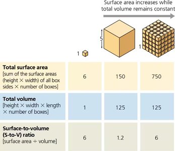
Q11. What happens to the surface area to volume ratio when cell volume increases? Use the figure and table above to determine how the volume of a cell changes compared to the surface area as the diameter increases. Which one increases the fastest or the most? How does the surface area to volume ratio change in the larger cell?
Background
Topic: Surface Area to Volume Ratio
This question tests your understanding of how cell size affects the efficiency of transport and metabolism.
Key Formula:
Step-by-Step Guidance
Observe how both surface area and volume change as the cell (cube) gets larger.
Calculate the surface area and volume for different cube sizes using the formulas above.
Compare how quickly each value increases as the side length increases.
Determine how the surface area to volume ratio changes as the cell grows.
Try solving on your own before revealing the answer!
Final Answer:
As cell size increases, volume increases faster than surface area, causing the surface area to volume ratio to decrease. This limits the efficiency of transport across the membrane in larger cells.
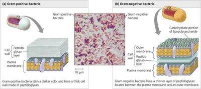
Q17. Using the figure above, describe the structural differences in gram positive vs gram negative bacteria.
Background
Topic: Prokaryotic Cell Walls and Antibiotics
This question tests your ability to distinguish between gram-positive and gram-negative bacteria based on cell wall structure.
Key Terms:
Peptidoglycan: Main component of bacterial cell walls.
Gram-positive: Thick peptidoglycan layer, no outer membrane.
Gram-negative: Thin peptidoglycan layer, outer membrane present.
Step-by-Step Guidance
Identify the main structural features of gram-positive bacteria (thick peptidoglycan, no outer membrane).
Identify the main structural features of gram-negative bacteria (thin peptidoglycan, outer membrane).
Explain how these differences affect staining and antibiotic susceptibility.
Relate these features to the effectiveness of antibiotics like penicillin.
Try solving on your own before revealing the answer!
Final Answer:
Gram-positive bacteria have a thick peptidoglycan layer and stain darkly; gram-negative bacteria have a thin peptidoglycan layer and an additional outer membrane, making them less susceptible to certain antibiotics.
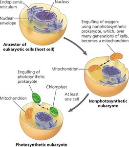
Q25. Describe the endosymbiosis theory and how the current eukaryotic cell may have derived with early origins with a prokaryotic cell. Use the diagram below in your description.
Background
Topic: Endosymbiosis Theory
This question tests your understanding of the evolutionary origin of mitochondria and chloroplasts in eukaryotic cells.
Key Terms:
Endosymbiosis: Theory that certain organelles originated as free-living prokaryotes engulfed by ancestral eukaryotic cells.
Mitochondria: Organelle for energy production, thought to have originated from aerobic bacteria.
Chloroplasts: Organelle for photosynthesis, thought to have originated from photosynthetic bacteria.
Step-by-Step Guidance
Describe the process of one cell engulfing another and forming a symbiotic relationship.
Explain how mitochondria and chloroplasts are believed to have originated from this process.
Discuss the evidence supporting this theory (e.g., double membranes, own DNA).
Relate the theory to the diversity of eukaryotic cells today.
Try solving on your own before revealing the answer!
Final Answer:
The endosymbiosis theory proposes that mitochondria and chloroplasts originated as free-living prokaryotes engulfed by ancestral eukaryotic cells, leading to a symbiotic relationship. Evidence includes their own DNA, double membranes, and similarities to bacteria.
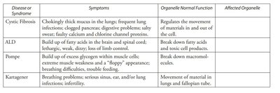
Q50. What organelles or cell structures are likely malfunctioning in these diseases?
Background
Topic: Organelle Function and Disease
This question tests your ability to connect symptoms of diseases to the malfunction of specific organelles.
Key Terms:
Cystic Fibrosis: Involves malfunction of membrane proteins.
ALD: Involves peroxisomes (fatty acid breakdown).
Pompe: Involves lysosomes (glycogen breakdown).
Kartagener: Involves cytoskeleton (cilia/flagella movement).
Step-by-Step Guidance
Read the symptoms and normal organelle function for each disease.
Match the symptoms to the organelle responsible (e.g., movement issues to cytoskeleton, digestion issues to lysosomes).
Consider how malfunction of each organelle leads to the observed symptoms.
Fill in the affected organelle for each disease in the table.
Try solving on your own before revealing the answer!
Final Answer:
Cystic Fibrosis: Plasma membrane (channel proteins); ALD: Peroxisomes; Pompe: Lysosomes; Kartagener: Cytoskeleton (cilia/flagella).
