 뒤로
뒤로Human Tissues: Structure, Function, and Classification
스터디 가이드 - 스마트 노트
 자료에 맞춘 맞춤형 노트, 핵심 정의, 예시, 맥락을 확장해 제공합니다.
Levels of Organization in Multicellular Organisms
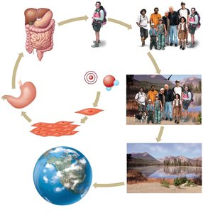
Biological Hierarchy
Multicellular organisms are organized into a hierarchy of increasing complexity, allowing for specialized functions and efficient life processes.
Cells: Basic unit of life, specialized for various functions.
Tissues: Groups of similar cells performing a common function.
Organs: Structures composed of multiple tissue types working together.
Organ Systems: Groups of organs that perform related functions.
Organism: An individual living entity.
Population, Community, Ecosystem, Biosphere: Higher levels of biological organization beyond the individual.
Major Tissue Types in the Human Body
Overview of Tissue Types
Tissues are groups of specialized cells with similar structure and function. The four major tissue types in humans are:
Epithelial Tissue
Connective Tissue
Muscle Tissue
Nervous Tissue
Epithelial Tissue
Structure and Function
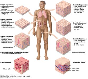
Epithelial tissues are sheets of cells that cover body surfaces, line cavities, and form glands. They serve as protective barriers, reduce friction, absorb nutrients, and secrete products.
Locations: Skin, lining of mouth, digestive tract, lungs, bladder, blood vessels, kidney tubules.
Functions: Protection, absorption, secretion, and sensation.
Specialization: Glandular epithelia form exocrine (secrete into ducts) and endocrine (secrete into blood) glands.
Classification of Epithelial Tissue
Epithelial tissues are classified by cell shape and number of layers:
By Shape:
Squamous: Flat, plate-like cells
Cuboidal: Cube-shaped cells
Columnar: Tall, column-shaped cells (may include goblet cells for mucus secretion)
By Layers:
Simple: Single layer
Stratified: Multiple layers
Basement Membrane and Cell Junctions
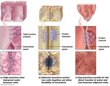
The basement membrane is a non-cellular layer beneath epithelial cells, providing support and anchoring to underlying connective tissue. Epithelial cells are held together by specialized junctions:
Tight Junctions: Form leak-proof seals between cells.
Adhesion (Anchoring) Junctions: Allow flexibility and hold cells together.
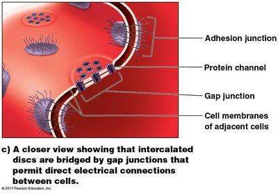
Gap Junctions: Permit direct transfer of water and ions between adjacent cells.
Connective Tissue
General Features and Functions
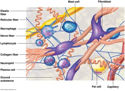
Connective tissues support, connect, and protect other tissues and organs. They have relatively few living cells embedded in an extracellular matrix composed of fibers and ground substance.
Functions: Support, binding, fat storage, blood cell production, defense.
Main Components: Cells (e.g., fibroblasts, adipocytes), fibers (collagen, elastic, reticular), ground substance.
Fibrous Connective Tissue
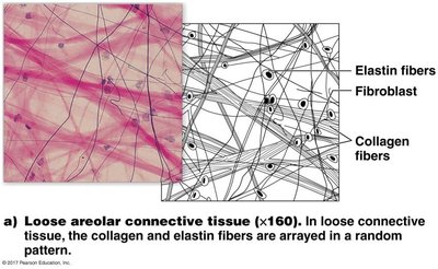
Loose (Areolar) Connective Tissue: Few collagen and elastic fibers in a random arrangement; provides flexibility and moderate strength; surrounds organs, blood vessels, and muscles.
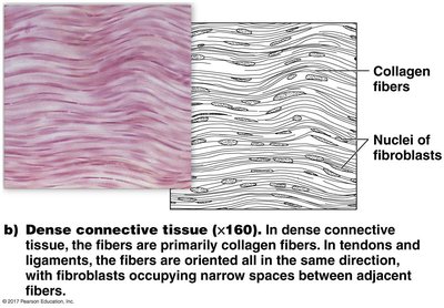
Dense Connective Tissue: Many collagen fibers aligned in one direction; very strong; found in tendons, ligaments, and lower layers of skin; heals slowly due to low cell and nutrient supply.
Elastic Connective Tissue: High proportion of elastic fibers; stretches and recoils easily; found in organs that change shape (e.g., bladder, stomach, vocal cords).
Reticular (Lymphoid) Connective Tissue: Thin, branched reticular fibers; forms internal framework for soft organs (e.g., liver, spleen, lymph nodes).
Specialized Connective Tissue
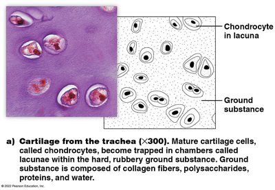
Cartilage: Flexible, supportive tissue with chondrocytes in lacunae; no blood vessels; found in joints, nose, ears, and between vertebrae.
Bone: Hard matrix with mineral deposits (Ca2+, PO43-); supports and protects; heals faster than cartilage due to blood supply.
Blood: Fluid matrix (plasma) with red cells (O2/CO2 transport), white cells (defense), and platelets (clotting).
Adipose Tissue: Specialized for fat storage in adipocytes; insulates and protects organs; number of adipocytes is genetically determined.
Muscle Tissue
General Features
Muscle tissues consist of specialized cells (fibers) that contract to produce movement. Contraction is due to interaction of proteins within the cytoplasm.
Three Types: Skeletal, Cardiac, Smooth
Skeletal Muscle
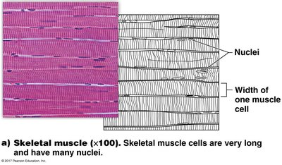
Function: Moves the body by contracting and pulling on bones via tendons.
Structure: Long, cylindrical, multinucleated fibers aligned in parallel; voluntary control.
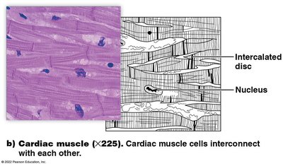
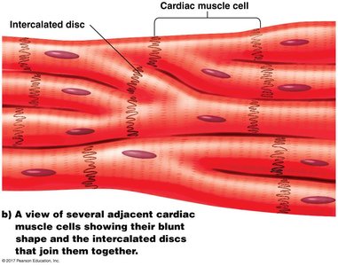
Cardiac Muscle
Function: Pumps blood through the heart and body.
Structure: Short, branched cells with one nucleus; connected by intercalated discs containing gap junctions for coordinated contraction; involuntary control.
Smooth Muscle
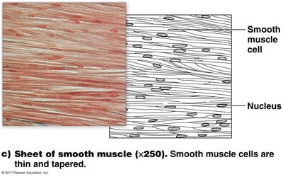
Function: Controls diameter of blood vessels, moves contents through hollow organs (e.g., digestive tract, bladder).
Structure: Small, spindle-shaped cells with one nucleus; involuntary control; cells contract together via gap junctions.
Comparison of Muscle Types
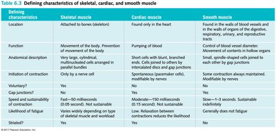
The three muscle types differ in location, structure, function, and control. The table below summarizes these differences:
Defining characteristics | Skeletal muscle | Cardiac muscle | Smooth muscle |
|---|---|---|---|
Location | Attached to bones (skeleton) | Found only in the heart | Walls of blood vessels and hollow organs |
Function | Movement of body, prevention of movement | Pumping of blood | Control diameter, move contents |
Anatomical description | Long, cylindrical, multinucleated, parallel bundles | Short, branched, single nucleus, intercalated discs | Small, spindle-shaped, single nucleus |
Initiation of contraction | Only by a nerve cell | Spontaneous (pacemaker), modulated by nerves | Some spontaneous, modulated by nerves/hormones |
Voluntary? | Yes | No | No |
Gap junctions? | No | Yes | Yes |
Speed and sustainability | Fast (10–100 ms), not sustainable | Moderate (50–150 ms), not sustainable | Slow (1–3 s), sustainable |
Likelihood of fatigue | High, depending on type | Low, relaxation between contractions reduces fatigue | Generally does not fatigue |
Striations visible? | Yes | Yes | No |
Nervous Tissue
Structure and Function
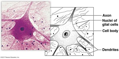
Nervous tissue contains specialized cells for generating and transmitting electrical impulses, allowing communication throughout the body. It is found in the brain, spinal cord, and nerves.
Two Main Cell Types:
Neurons: Conduct electrical impulses.
Glial Cells: Support, protect, and nourish neurons; do not conduct impulses.
Parts of a Neuron
Cell Body: Contains nucleus and organelles.
Dendrites: Receive signals from other cells.
Axon: Conducts electrical impulses away from the cell body.
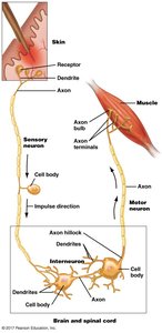
Types of Neurons
Sensory Neurons: Receive stimuli and transmit information to the central nervous system (CNS).
Interneurons: Integrate information within the CNS.
Motor Neurons: Transmit signals from the CNS to muscles and glands for response.
Glial Cells
Provide physical support and protection for neurons.
Maintain chemical and nutrient balance around neurons.
Comprise about 80% of cells in the nervous system.
Do not generate electrical impulses.
Additional info: The study of tissues is called histology. Understanding tissue structure and function is foundational for advanced studies in anatomy, physiology, and pathology.
