 뒤로
뒤로Introduction to Cells and Their Structures
스터디 가이드 - 스마트 노트
 자료에 맞춘 맞춤형 노트, 핵심 정의, 예시, 맥락을 확장해 제공합니다.
Introduction to Cells and Their Structures
Cell Theory
The cell theory is a foundational concept in biology, describing the properties and significance of cells in living organisms.
All living things are composed of cells and cell products.
The cell is the smallest unit that exhibits all the characteristics of life.
All cells arise only from preexisting cells.
These principles remain central to modern biology and guide our understanding of life at the cellular level.
Classification of Cells
Cells are classified based on their internal organization and complexity into two main types: prokaryotic and eukaryotic cells.
Prokaryotic Cells: Simpler structure, lack membrane-bound organelles, DNA is located in a region called the nucleoid, surrounded by a plasma membrane and often a rigid cell wall. Examples: Bacteria, blue-green algae.
Eukaryotic Cells: More complex, contain a true nucleus (membrane-bound DNA region), cytoplasm with organelles, and a plasma membrane. Examples: Animals, plants.
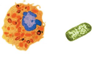
Figure: Comparison of eukaryotic (left) and prokaryotic (right) cell structures.
Structure Reflects Function
Although all cells share basic functions such as gathering raw materials, excreting wastes, synthesizing macromolecules, and reproducing, their structures vary to suit specialized roles:
Muscle cells: Abundant organelles for energy production.
Nerve cells: Long and thin to transmit signals over distances.
Red blood cells: Round and flexible to move through capillaries.
Each eukaryotic cell type is specialized for its function.
Cell Size and Efficiency
Cells remain small to maximize efficiency in exchanging materials with their environment. As a cell grows, its volume increases faster than its surface area, limiting the rate at which materials can cross the plasma membrane.
Surface area-to-volume ratio: Smaller cells have a higher ratio, allowing more efficient transport of materials.
Microvilli: Microscopic projections that increase surface area, commonly found in the digestive tract and kidney tubules.
Example: Eight small cells have a greater total surface area than one large cell of the same total volume, enhancing material exchange.
Microscopes
Microscopes are essential tools for studying cells due to their small size. There are three main types:
Light Microscope: Uses light to magnify specimens up to 1000x; suitable for viewing live cells and general structures.
Transmission Electron Microscope (TEM): Uses electrons to produce detailed 2D images of internal cell structures; magnifies up to 100,000x.
Scanning Electron Microscope (SEM): Uses electrons to create 3D images of cell surfaces; magnifies up to 100,000x.
Internal Cell Structures and Functions
The Nucleus
The nucleus is the control center of eukaryotic cells, containing most of the cell's genetic material.
Nuclear membrane: Double phospholipid bilayer that encloses DNA.
Nucleolus: Dense region where ribosomal components are formed.
Nuclear pores: Allow selective movement of materials (e.g., ribosomes, proteins, RNA) in and out of the nucleus; DNA remains inside.
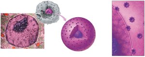
Figure: Structure of the nucleus, showing the nucleolus and nuclear pores.
Ribosomes
Ribosomes are the sites of protein synthesis, assembling amino acids into polypeptide chains according to genetic instructions.
Formed from RNA and proteins in the nucleolus.
Can be free in the cytoplasm or attached to the endoplasmic reticulum.
Endomembrane System
The endomembrane system includes several interconnected organelles responsible for the synthesis, modification, and transport of cellular materials:
Rough Endoplasmic Reticulum (RER): Studded with ribosomes; synthesizes proteins.
Smooth Endoplasmic Reticulum (SER): Lacks ribosomes; synthesizes lipids and hormones, packages proteins and lipids into vesicles.
Golgi Apparatus: Modifies, sorts, and packages proteins and lipids for delivery.
Specialized Vesicles: Transport materials within and out of the cell.
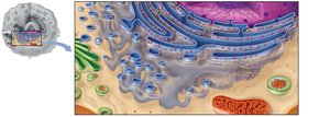
Figure: Structure of the endoplasmic reticulum, showing rough and smooth regions.
Golgi Apparatus
The Golgi apparatus receives vesicles from the ER, refines and labels proteins and lipids, and packages them for transport to their final destinations.
Enzymes within the Golgi modify products.
Sorts and ships products via vesicles.
Lipid synthesis also occurs here.
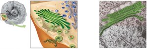
Figure: Structure and function of the Golgi apparatus.
Vesicles
Vesicles are small, membrane-bound sacs that transport and store substances within a cell.
Secretory vesicles: Export products out of the cell.
Endocytotic vesicles: Bring external materials into the cell.
Peroxisomes: Contain enzymes to break down toxic wastes.
Lysosomes: Contain digestive enzymes to break down bacteria, debris, and damaged organelles.
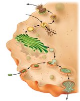
Figure: Lysosomes and peroxisomes forming from the Golgi apparatus.
Mitochondria
Mitochondria are the powerhouses of the cell, converting nutrients into ATP through cellular respiration.
Double membrane structure: smooth outer membrane, highly folded inner membrane (cristae).
Contain their own DNA and ribosomes.
Number varies with cell energy needs (e.g., muscle cells have many mitochondria).
Cytoskeleton
The cytoskeleton provides structural support, maintains cell shape, and anchors organelles.
Microtubules: Hollow tubes for support and transport.
Microfilaments: Thin fibers for movement and shape.
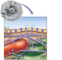
Figure: Cytoskeleton structure and its role in supporting the cell.
Cilia and Flagella
Both cilia and flagella are made of microtubules and are involved in movement:
Cilia: Short, numerous projections that move materials along the cell surface (e.g., in respiratory tract).
Flagella: Longer, fewer in number, used for cell movement (e.g., sperm cells).
Centrioles
Centrioles are short, rod-like structures near the nucleus, essential for cell division. They help organize the alignment and separation of genetic material during mitosis and meiosis.
Summary Table: Major Eukaryotic Cell Structures and Functions
Structure | Main Function |
|---|---|
Plasma membrane | Controls movement of materials into and out of cell |
Nucleus | Information center; contains DNA |
Ribosomes | Site of protein synthesis |
Rough ER | Protein synthesis and processing |
Smooth ER | Lipid and hormone synthesis; detoxification |
Golgi apparatus | Modifies, sorts, and ships macromolecules |
Lysosomes | Digestion of macromolecules and debris |
Peroxisomes | Breakdown of toxic substances |
Mitochondria | ATP (energy) production |
Cytoskeleton | Structural support and movement |
Centrioles | Cell division |
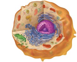
Figure: Diagram of a eukaryotic cell with labeled organelles and their functions.
