 뒤로
뒤로Membrane Structure and Function: Study Guide
스터디 가이드 - 스마트 노트
 자료에 맞춘 맞춤형 노트, 핵심 정의, 예시, 맥락을 확장해 제공합니다.
Membrane Structure and Function
Overview
The plasma membrane is a dynamic structure that separates the interior of the cell from its external environment. It regulates the movement of substances in and out of the cell, maintaining homeostasis and enabling communication and transport.
Fluid Mosaic Model of Membrane Structure
Components of the Membrane
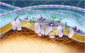
The fluid mosaic model describes the plasma membrane as a flexible layer made of lipid molecules interspersed with large protein molecules that act as channels through which other molecules enter and leave the cell.
Phospholipids: Amphipathic molecules with hydrophilic heads and hydrophobic tails, forming a bilayer.
Cholesterol: Stabilizes membrane fluidity across temperature changes.
Proteins: Integral (span the membrane) and peripheral (attached to the surface).
Carbohydrates: Attached to lipids (glycolipids) or proteins (glycoproteins), important for cell recognition.
Cytoskeleton and ECM: Provide structural support and facilitate communication.
Membrane Fluidity
Decreasing temperature: Reduces fluidity as phospholipids pack more tightly.
Unsaturated hydrocarbon chains: Increase fluidity due to kinks in tails preventing tight packing.
Cholesterol: Acts as a fluidity buffer, preventing extremes in fluidity.
Saturated hydrocarbon tails: Decrease fluidity by allowing tighter packing.
Membrane Proteins
Integral proteins: Penetrate the hydrophobic core; often function as transport channels or receptors.
Peripheral proteins: Loosely bound to the membrane surface; often involved in signaling or maintaining cell shape.
Functions of Membrane Proteins
Transport: Move substances across the membrane.
Enzymatic activity: Catalyze reactions at the membrane surface.
Signal transduction: Relay signals from outside to inside the cell.
Cell-cell recognition: Allow cells to identify each other.
Intercellular joining: Connect adjacent cells.
Attachment to cytoskeleton and ECM: Maintain cell shape and stabilize membrane proteins.
Membrane Carbohydrates
Glycolipids: Carbohydrates covalently bonded to lipids.
Glycoproteins: Carbohydrates covalently bonded to proteins.
Selective Permeability of Membranes
How Structure Determines Permeability
The lipid bilayer is selectively permeable, allowing some substances to cross more easily than others. Small, nonpolar molecules pass freely, while ions and large polar molecules require transport proteins.
Transport Proteins
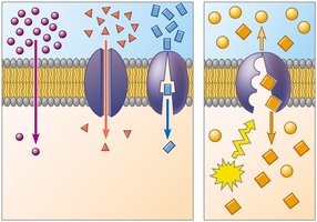
Channel proteins: Provide corridors for specific molecules or ions to cross.
Carrier proteins: Bind to molecules and change shape to shuttle them across the membrane.
Specificity: Transport proteins are specific for the substance they move (e.g., glucose transporters).
Aquaporins: Channel proteins that facilitate water transport.
Types of Membrane Transport
Passive Transport
Passive transport does not require energy and includes diffusion, osmosis, and facilitated diffusion.
Diffusion: Movement of molecules from high to low concentration.
Concentration gradient: The difference in concentration across a space.
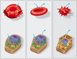
Osmosis: Diffusion of water across a selectively permeable membrane.
Isotonic: Equal solute concentration inside and outside the cell.
Hypertonic: Higher solute concentration outside the cell; cell loses water.
Hypotonic: Lower solute concentration outside the cell; cell gains water.
Turgid: Firm plant cell in hypotonic solution.
Flaccid: Limp plant cell in isotonic solution.
Plasmolysis: Plant cell membrane pulls away from the wall in hypertonic solution.
Facilitated Diffusion
Facilitated diffusion is passive transport aided by proteins. It includes channel proteins (e.g., ion channels) and carrier proteins (e.g., glucose transporter).
Active Transport
Mechanism and Example
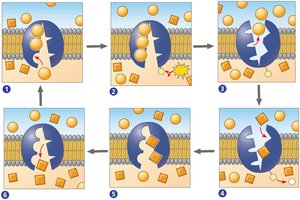
Active transport moves substances against their concentration gradients using energy (usually ATP) and carrier proteins (pumps).
Sodium–potassium pump: Moves 3 Na+ out and 2 K+ into the cell, maintaining electrochemical gradients.
ATP: Provides energy by transferring a phosphate group to the transport protein.
Membrane Potential and Electrochemical Gradient
Membrane potential: Voltage across the membrane, usually negative inside the cell.
Electrochemical gradient: Combination of concentration and electrical gradients driving ion movement.
Cotransport
Cotransport uses the energy of one molecule moving down its gradient to drive another molecule against its gradient (e.g., glucose-sodium cotransport in intestines).
Bulk Transport
Mechanisms
Exocytosis: Vesicles fuse with the membrane to release contents outside the cell (e.g., neurotransmitter release).
Endocytosis: Cell takes in materials by forming vesicles from the membrane.
Phagocytosis: "Cell eating"; engulfing large particles.
Pinocytosis: "Cell drinking"; uptake of extracellular fluid.
Receptor-mediated endocytosis: Specific uptake of molecules via receptor proteins.
Application: Osmosis and Diffusion in Cells
Experimental Example
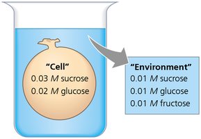
Consider a cell with 0.03 M sucrose and 0.02 M glucose in an environment with 0.01 M sucrose, 0.01 M glucose, and 0.01 M fructose. The movement of water and solutes will depend on their concentration gradients and membrane permeability.
Summary Table: Types of Membrane Transport
Type of Transport | Energy Required? | Direction | Example |
|---|---|---|---|
Simple Diffusion | No | Down gradient | O2, CO2 |
Facilitated Diffusion (Channel) | No | Down gradient | Ion channels |
Facilitated Diffusion (Carrier) | No | Down gradient | Glucose transporter |
Active Transport | Yes (ATP) | Against gradient | Sodium-potassium pump |
Bulk Transport | Yes | In or out | Exocytosis, endocytosis |
Additional info: This guide expands on the provided notes with definitions, examples, and a summary table for clarity and completeness.
