 뒤로
뒤로The Cell Cycle and Cell Division: Structure, Function, and Regulation
스터디 가이드 - 스마트 노트
 자료에 맞춘 맞춤형 노트, 핵심 정의, 예시, 맥락을 확장해 제공합니다.
The Key Roles of Cell Division
Introduction to Cell Division
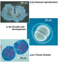
Cell division is a fundamental process that distinguishes living organisms from nonliving matter. It ensures the continuity of life by enabling organisms to reproduce, grow, and repair tissues. In unicellular organisms, cell division results in the reproduction of the entire organism, while in multicellular eukaryotes, it is essential for development, growth, and tissue renewal.
Asexual reproduction: Single-celled organisms reproduce by dividing into two identical cells.
Growth and development: Multicellular organisms grow by increasing their cell number through division.
Tissue renewal: Damaged or old cells are replaced by new cells formed through division.
Cellular Organization of Genetic Material
Chromosomes and Chromatin
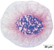
All the DNA in a cell constitutes its genome. In prokaryotes, the genome is typically a single DNA molecule, while in eukaryotes, it consists of multiple DNA molecules packaged into chromosomes. Eukaryotic chromosomes are composed of chromatin, a complex of DNA and protein that condenses during cell division.
Somatic cells: Nonreproductive cells with two sets of chromosomes.
Gametes: Reproductive cells (sperm and eggs) with half as many chromosomes as somatic cells.
Chromosome Duplication and Distribution
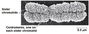
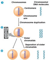
Before cell division, chromosomes are duplicated, resulting in two sister chromatids joined at a region called the centromere. During division, sister chromatids separate and are distributed into two daughter cells, ensuring genetic continuity.
Sister chromatids: Identical copies of a chromosome connected by cohesins.
Centromere: The region where sister chromatids are most closely attached.
The Cell Cycle
Phases of the Cell Cycle
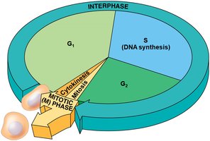
The cell cycle is the ordered sequence of events in the life of a cell, from its formation to its own division. It consists of two main phases: interphase and the mitotic (M) phase.
Interphase: Accounts for about 90% of the cell cycle and includes three subphases:
G1 phase (first gap): Cell growth.
S phase (synthesis): DNA replication.
G2 phase (second gap): Preparation for mitosis.
Mitotic (M) phase: Includes mitosis (division of the nucleus) and cytokinesis (division of the cytoplasm).
Mitosis: The Process of Nuclear Division
Stages of Mitosis
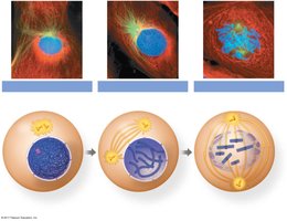
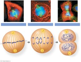
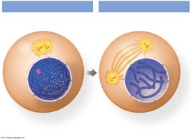
Mitosis is conventionally divided into five stages, each characterized by specific events that ensure accurate chromosome segregation:
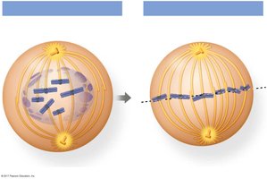
Prophase: Chromosomes condense, and the mitotic spindle begins to form.
Prometaphase: Nuclear envelope fragments, and spindle microtubules attach to kinetochores.
Metaphase: Chromosomes align at the metaphase plate.
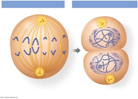
Anaphase: Sister chromatids separate and move toward opposite poles.
Telophase: Nuclear envelopes reform around the chromosomes, which decondense.
The Mitotic Spindle and Chromosome Movement
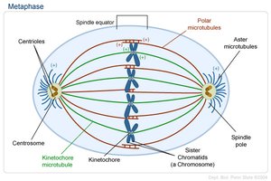
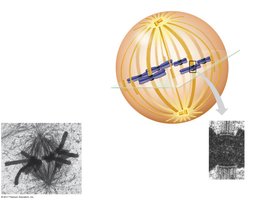
The mitotic spindle is a structure made of microtubules that orchestrates the movement of chromosomes during mitosis. In animal cells, spindle assembly begins at the centrosomes, which replicate and migrate to opposite poles. Microtubules attach to kinetochores on chromosomes, facilitating their movement.
Aster: A radial array of short microtubules extending from each centrosome.
Metaphase plate: An imaginary plane where chromosomes align during metaphase.
Mechanisms of Chromosome Movement
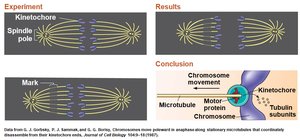
During anaphase, cohesins are cleaved by the enzyme separase, allowing sister chromatids to separate. Chromatids move toward opposite poles, primarily due to the action of motor proteins and the depolymerization of microtubules at the kinetochore ends—a process known as the "Pac-man" mechanism.
Cytokinesis: Division of the Cytoplasm
Mechanisms of Cytokinesis
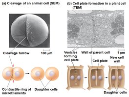
Cytokinesis is the process by which the cytoplasm divides, resulting in two daughter cells. In animal cells, this occurs via cleavage, forming a cleavage furrow. In plant cells, a cell plate forms, eventually developing into a new cell wall.
Binary Fission in Prokaryotes
Prokaryotic Cell Division
Prokaryotes such as bacteria reproduce by binary fission. The chromosome replicates, and the two copies move to opposite ends of the cell. The plasma membrane pinches inward, dividing the cell into two genetically identical daughter cells.
Evolutionary Perspective
Mitosis likely evolved from binary fission, as certain protists exhibit intermediate forms of cell division.
Regulation of the Eukaryotic Cell Cycle
The Cell Cycle Control System
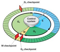
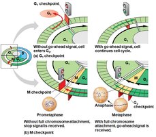
The eukaryotic cell cycle is regulated by a molecular control system, which ensures that cell division occurs only when appropriate. This system operates like a clock, with specific checkpoints (G1, G2, and M) where the cycle can be halted until certain conditions are met.
Cyclins and Cyclin-Dependent Kinases (Cdks)
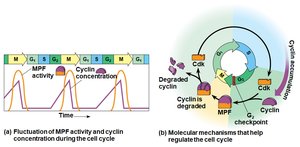
Two key types of regulatory proteins control the cell cycle: cyclins and cyclin-dependent kinases (Cdks). The activity of Cdks fluctuates with the concentration of their cyclin partners. The maturation-promoting factor (MPF) is a cyclin-Cdk complex that triggers passage through the G2 checkpoint into mitosis.
Checkpoints and Signals
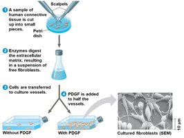
Checkpoints are regulated by internal and external signals. For example, cells will not begin anaphase until all chromosomes are properly attached to the spindle at the metaphase plate. External signals include growth factors such as platelet-derived growth factor (PDGF), which stimulate cell division.
Loss of Cell Cycle Control and Cancer
Cancer and Uncontrolled Cell Division
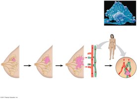
Cancer cells do not respond to normal regulatory signals. They may produce their own growth factors, convey signals without external stimuli, or have abnormal control systems. Cells that divide indefinitely undergo transformation and may form tumors. Tumors can be benign (localized) or malignant (invasive and capable of metastasis).
Modern Cancer Treatments
Advances in understanding cell cycle regulation have led to targeted cancer therapies. Personalized treatments are increasingly possible due to the ability to sequence tumor DNA and identify specific molecular abnormalities.
