 뒤로
뒤로The Molecular Basis of Inheritance: DNA Structure and Replication
스터디 가이드 - 스마트 노트
 자료에 맞춘 맞춤형 노트, 핵심 정의, 예시, 맥락을 확장해 제공합니다.
Chapter 16 – The Molecular Basis of Inheritance
The Search for the Genetic Material
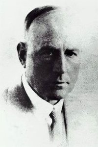
Early 20th-century biologists sought to identify the molecules responsible for inheritance. Chromosomes, composed of DNA and protein, were the main candidates. The discovery of DNA's genetic role began with Frederick Griffith's experiments in 1928, which demonstrated the phenomenon of transformation in bacteria.
Evidence That Viral DNA Can Program Cells
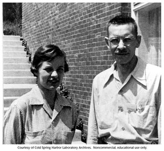
Viruses, which can contain either DNA or RNA enclosed in a protein coat, provided further evidence for DNA as the genetic material. Bacteriophages (phages) are viruses that infect bacteria and have been instrumental in molecular genetics research. In 1952, Alfred Hershey and Martha Chase used the T2 phage to show that DNA, not protein, is the genetic material injected into E. coli during infection.
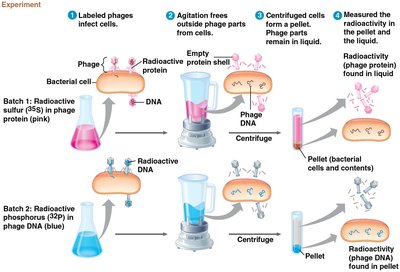
Key Point: Only DNA entered the bacterial cells and directed viral reproduction, confirming DNA as the genetic material.
Example: The Hershey-Chase experiment used radioactive isotopes to label DNA and protein, tracking which component entered the cell.
Chargaff’s Rules and DNA Composition
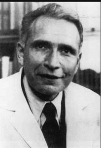
Erwin Chargaff discovered that DNA composition varies between species and that the amount of adenine (A) equals thymine (T), while guanine (G) equals cytosine (C). These findings, known as Chargaff’s rules, provided clues to DNA’s structure.
Key Point: Base pairing regularity (A = T, G = C) is universal among organisms.
Building a Structural Model of DNA
X-ray Crystallography and the Double Helix
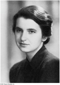
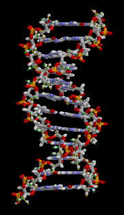
Maurice Wilkins and Rosalind Franklin used X-ray crystallography to study DNA’s molecular structure. Franklin’s famous X-ray diffraction image enabled James Watson and Francis Crick to deduce that DNA is helical, with a uniform width and repeating structure, suggesting a double helix composed of two antiparallel strands.
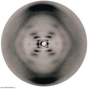
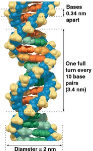
Key Point: The double helix consists of two sugar-phosphate backbones on the outside, with nitrogenous bases paired in the interior.
Example: The helical structure repeats every 3.4 nm, with bases stacked 0.34 nm apart.
Base Pairing and Antiparallel Strands
Watson and Crick’s model showed that DNA strands run in opposite directions (antiparallel). Base pairing is specific: adenine pairs with thymine (A–T) and guanine pairs with cytosine (G–C), forming hydrogen bonds. Pairing a purine with a pyrimidine maintains a uniform helix width, consistent with X-ray data.
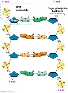
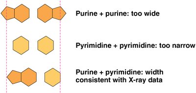
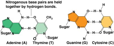
Key Point: The specificity of base pairing explains Chargaff’s rules and ensures accurate DNA replication.
DNA Replication
The Basic Principle: Base Pairing to a Template Strand
DNA replication is the process by which DNA makes an exact copy of itself. Each strand serves as a template for the formation of a new complementary strand, resulting in two identical DNA molecules.
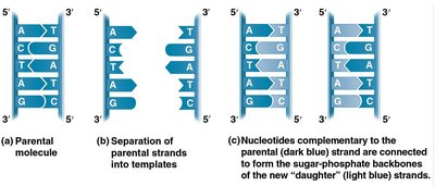
Key Point: The semiconservative model predicts that each daughter DNA molecule consists of one parental and one new strand.
Experimental Evidence for the Semiconservative Model
Matthew Meselson and Franklin Stahl’s experiments using isotopic labeling of DNA confirmed the semiconservative model of replication.
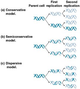
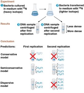
Key Point: After one round of replication, DNA molecules had intermediate density, ruling out the conservative model.
DNA Replication: A Closer Look
DNA replication is a highly accurate and rapid process involving many enzymes and proteins. The process is fundamentally similar in prokaryotes and eukaryotes.
Origins of Replication
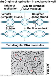
Replication begins at specific sites called origins of replication, where the DNA strands separate to form replication bubbles. Eukaryotic chromosomes have multiple origins, while prokaryotic chromosomes typically have one.
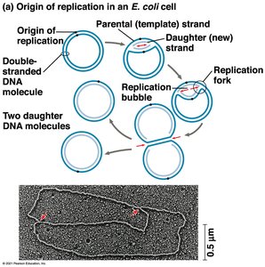
Key Point: Replication proceeds in both directions from each origin until the entire molecule is copied.
Enzymes and Proteins in DNA Replication
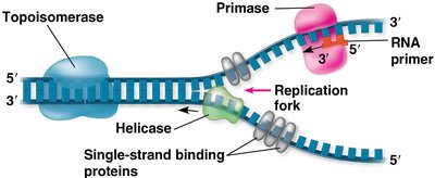
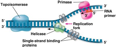
Several key enzymes and proteins are involved in DNA replication:
Helicase: Unwinds the DNA double helix at the replication fork.
Single-strand binding proteins: Stabilize unwound DNA strands.
Topoisomerase: Relieves strain caused by unwinding by breaking and rejoining DNA strands.
Primase: Synthesizes a short RNA primer to provide a starting point for DNA polymerase.
Synthesizing a New DNA Strand
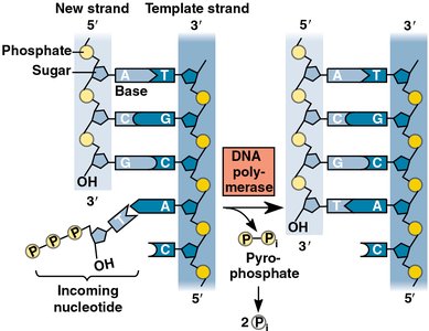
DNA polymerases catalyze the addition of nucleotides to the growing DNA strand, starting from the 3′ end of the RNA primer. Each nucleotide added is a nucleoside triphosphate, and the reaction releases pyrophosphate.
Key Point: DNA polymerases can only add nucleotides to the 3′ end, so DNA synthesis always proceeds in the 5′ → 3′ direction.
Leading and Lagging Strands
Because DNA polymerase can only synthesize in one direction, replication is continuous on the leading strand and discontinuous on the lagging strand. The lagging strand is synthesized in short segments called Okazaki fragments, which are joined by DNA ligase.
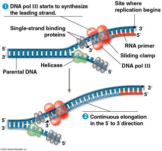
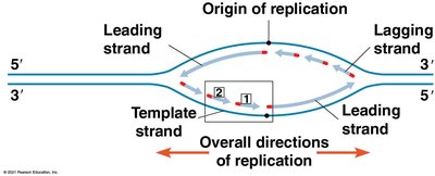
Key Point: The antiparallel structure of DNA results in different replication mechanisms for the two strands.
The DNA Replication Complex
The proteins involved in DNA replication form a large complex, sometimes called the DNA replication machine. This complex may be stationary, with DNA moving through it during replication.
Proofreading and Repairing DNA
DNA polymerases proofread newly synthesized DNA, correcting errors. Additional repair mechanisms, such as mismatch repair and nucleotide excision repair, fix errors and damage caused by environmental factors.
Key Point: The error rate after proofreading and repair is extremely low, ensuring genetic fidelity.
Evolutionary Significance of DNA Mutations
Although rare, mutations that escape repair can be inherited. These mutations are the source of genetic variation, which is essential for evolution by natural selection.
Replicating the Ends of DNA Molecules
In eukaryotes, the replication machinery cannot fully replicate the 5′ ends of linear DNA, leading to progressive shortening. Telomeres, repetitive nucleotide sequences at chromosome ends, protect genes from erosion. The enzyme telomerase extends telomeres in germ cells, and its activity is linked to aging and cancer.
Chromosome Structure and DNA Packing
Bacterial and Eukaryotic Chromosomes
Bacterial chromosomes are circular DNA molecules with some associated proteins, found in the nucleoid region. Eukaryotic DNA is packaged with proteins into chromatin, which is organized into nucleosomes—DNA wrapped around histone protein cores. Chromatin further coils and condenses to form metaphase chromosomes during cell division.
Key Point: DNA packaging allows long DNA molecules to fit inside the cell nucleus and plays a role in gene regulation.
Feature | Bacterial Chromosome | Eukaryotic Chromosome |
|---|---|---|
Shape | Circular | Linear |
Location | Nucleoid | Nucleus |
Associated Proteins | Few | Many (histones) |
Packing Structure | Supercoiled | Chromatin, nucleosomes |
Additional info: Chromatin structure is dynamic and can be modified to regulate access to DNA for transcription, replication, and repair.
