 뒤로
뒤로Extracellular Matrix and Cell Adhesion: Structure, Function, and Clinical Relevance
스터디 가이드 - 스마트 노트
 자료에 맞춘 맞춤형 노트, 핵심 정의, 예시, 맥락을 확장해 제공합니다.
Extracellular Matrix (ECM) Overview
Structure and Functions of the ECM
The extracellular matrix (ECM) is a complex network of proteins and polysaccharides that provides structural and biochemical support to surrounding cells. Its composition and physical properties vary between tissues, ranging from the fluid ECM of blood to the spongy ECM of cartilage.
Structural support: Maintains tissue integrity and shape.
Cell adhesion: Mediates attachment of cells to the ECM and to each other.
Cell-cell communication: Facilitates signaling between cells.
Tissue organization: Guides cell migration and tissue patterning.
Growth factor reservoir: Stores and releases signaling molecules.
Dynamic remodeling: ECM is constantly remodeled during development, repair, and disease.
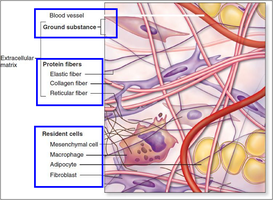
Major Components of the ECM
Fibrous Proteins
Fibrous proteins are the primary structural elements of the ECM, providing tensile strength and elasticity. The main types include collagen and elastin.
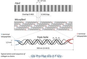
Collagen: The most abundant protein in the ECM, with at least 28 types (I–XXVIII). Forms triple-helical structures for tensile strength.
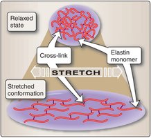
Elastin: Provides elasticity, allowing tissues such as skin, arteries, and lungs to stretch and recoil.
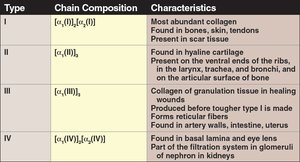
Collagen Types and Properties
Type | Chain Composition | Characteristics |
|---|---|---|
I | \( [\alpha_1(\mathrm{I})]_2 \alpha_2(\mathrm{I}) \) | Most abundant; found in bones, skin, tendons; present in scar tissue |
II | \( [\alpha_1(\mathrm{II})]_3 \) | Hyaline cartilage; ribs, larynx, trachea, bronchi, articular surfaces |
III | \( [\alpha_1(\mathrm{III})]_3 \) | Granulation tissue; reticular fibers; arteries, intestine, uterus |
IV | \( [\alpha_1(\mathrm{IV})]_2 \alpha_2(\mathrm{IV}) \) | Basal lamina, eye lens; filtration in kidney glomeruli |
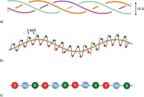
Collagen Structure
Collagen is characterized by a repeating Gly-X-Y amino acid sequence, where X is often proline and Y is often hydroxyproline. This sequence allows the formation of a tight triple helix, essential for its mechanical properties.
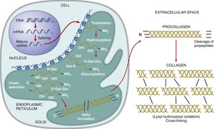
Collagen Synthesis and Assembly
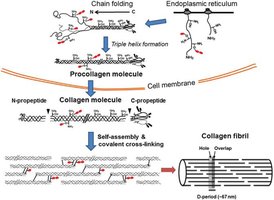
Collagen biosynthesis involves both intracellular and extracellular steps, including post-translational modifications, triple helix formation, secretion, and assembly into fibrils.
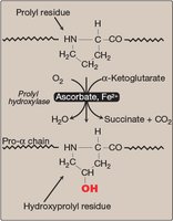
Hydroxylation: Proline and lysine residues are hydroxylated (requires vitamin C and Fe2+).
Glycosylation: Addition of sugars to hydroxylysine residues.
Triple helix formation: Three α-chains assemble into a procollagen triple helix.
Secretion and processing: Procollagen is secreted and terminal propeptides are cleaved.
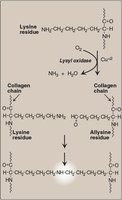
Crosslinking: Lysyl oxidase catalyzes covalent crosslinks for fibril stability.
Key Enzymes in Collagen Modification
Prolyl hydroxylase: Hydroxylates proline residues (requires ascorbate/vitamin C and Fe2+).
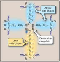
Lysyl oxidase: Catalyzes oxidative deamination of lysine, forming allysine for crosslinking (requires Cu2+).
Collagen in Disease: Scurvy
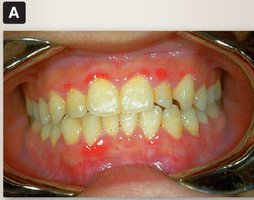
Vitamin C deficiency impairs proline hydroxylation, leading to defective collagen assembly. This results in fragile connective tissue, poor wound healing, and symptoms such as bleeding gums and tooth loss.
Type IV Collagen and Basement Membrane Structure
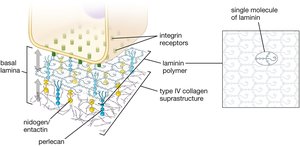
Type IV collagen forms a mesh-like network in basement membranes, unlike the rope-like fibrils of types I–III. Its assembly involves retention of NC1 and 7S domains, allowing lateral and end-to-end interactions for a planar scaffold.
Elastin
Elastin provides elasticity to tissues. Elastin monomers are crosslinked via desmosine bonds, allowing tissues to stretch and recoil.
Found in skin, arteries, lungs.
Crosslinking is essential for function.
Adhesive Glycoproteins
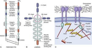
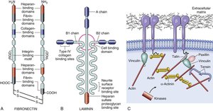
Adhesive proteins such as fibronectin and laminin connect ECM components and cells. They contain multiple binding domains for collagen, integrins, and proteoglycans.
Fibronectin: Dimeric, binds collagen, integrins, and proteoglycans.
Laminin: Heterotrimeric, binds collagen, cell surfaces, and proteoglycans; key in basement membranes.
Proteoglycans and Glycosaminoglycans (GAGs)
Glycosaminoglycans (GAGs)
GAGs are long, unbranched polysaccharides composed of repeating disaccharide units. They are highly negatively charged, attract water, and provide compressive strength and hydration to tissues.
Types: Hyaluronic acid, chondroitin sulfate, dermatan sulfate, keratan sulfate, heparan sulfate.
Functions: Space filling, water retention, resistance to compression.
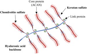
Proteoglycans
Proteoglycans consist of a core protein with covalently attached GAG chains. They are major ECM components in connective tissues such as bone, cartilage, and cornea.
Types: Aggrecan, decorin, perlecan, syndecan.
Functions: Hydration, compression resistance, growth factor binding.
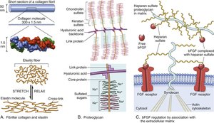
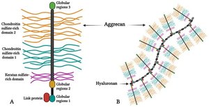
Proteoglycan Structure and Function
Proteoglycans have a "bottle brush" appearance due to their central protein core and radiating GAG chains. The negative charge of GAGs attracts Na+ ions and water, contributing to tissue hydration and resilience.
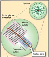
Cartilage and Synovial Fluid
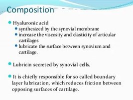
Cartilage is composed of chondrocytes embedded in a matrix of collagen, proteoglycans, and other proteins. Synovial fluid, rich in hyaluronic acid, lubricates joints and reduces friction.
ECM Dynamics and Remodeling
ECM Degradation and Remodeling
ECM components are constantly remodeled by enzymes such as matrix metalloproteinases (MMPs), ADAMs, and ADAMTS. This process is essential for wound repair, bone remodeling, and tissue homeostasis. Dysregulation can lead to fibrosis, cancer, or congenital defects.
Wound Healing and Provisional Matrix
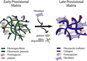
After injury, a provisional matrix forms, rich in fibrin and fibronectin, providing a scaffold for cell migration and tissue repair. Over time, this is replaced by a mature ECM containing collagen and proteoglycans.
Cell-ECM Interactions
Basement Membrane
The basement membrane is a specialized ECM structure composed of type IV collagen, laminin, nidogen, and perlecan. It serves as a barrier, maintains cell polarity, and regulates filtration (e.g., in the kidney glomerulus).
Integrins and Cell-ECM Adhesion
Integrins are transmembrane receptors that mediate cell-ECM adhesion and bidirectional signaling. They recognize ECM proteins and link the cytoskeleton to the ECM.
19 α and 8 β subunits form various integrin heterodimers.
Integrins participate in both inside-out and outside-in signaling.
Anchoring Junctions: Hemidesmosomes and Focal Adhesions
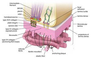
Hemidesmosomes: Link intermediate filaments to the ECM via integrins, providing stable adhesion to the basal lamina.
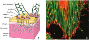
Focal adhesions: Connect actin cytoskeleton to the ECM, function in signaling, cell motility, and mechanosensation.
Integrin Signaling Cascade
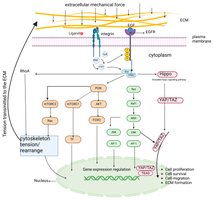
Integrin engagement with ECM proteins (e.g., fibronectin) triggers a signaling cascade involving FAK, Src, and the MAPK pathway, leading to changes in gene expression, proliferation, migration, and survival.
Cell-Cell Adhesion
Types of Cell-Cell Junctions
Anchoring junctions: Desmosomes and adherens junctions provide mechanical strength and connect cytoskeletons of adjacent cells.
Occluding junctions: Tight junctions (zonula occludens) form barriers to paracellular transport and maintain cell polarity.
Communicating junctions: Gap junctions allow passage of ions and small molecules for cell-cell communication.
Adhesion Molecules
Cadherins: Mediate calcium-dependent homotypic cell-cell adhesion; link to actin cytoskeleton.
Immunoglobulin superfamily: Mediate homotypic adhesion; roles in immunity and inflammation.
Selectins: Mediate heterotypic adhesion, especially in leukocyte-endothelial interactions.
Clinical Relevance: ECM and Adhesion in Disease
Genetic Disorders
Ehlers-Danlos Syndrome: Collagen defects cause joint hypermobility, skin hyperextensibility, and tissue fragility.
Marfan Syndrome: Fibrillin-1 mutations lead to aortic complications, skeletal abnormalities, and loss of tissue elasticity.
Menkes Disease: Copper deficiency impairs lysyl oxidase, leading to defective collagen crosslinking and connective tissue weakness.
Acquired Conditions
Fibrosis: Excessive ECM deposition causes organ dysfunction.
Osteoarthritis: ECM degradation in cartilage leads to joint pain and dysfunction; MMPs and ADAMTS enzymes are involved.
Cancer: ECM remodeling facilitates tumor invasion and metastasis; desmoplasia is the formation of dense fibrous tissue around tumors.
Pemphigus: Autoantibodies against desmoglein disrupt desmosomes, causing skin blistering.
Celiac Disease: Disruption of tight junctions increases intestinal permeability.
Cardiac Disease: Gap junction abnormalities can cause arrhythmias.
