 뒤로
뒤로Study Guide: The Macromolecules of the Cell – Protein Structure and Function
스터디 가이드 - 스마트 노트
 자료에 맞춘 맞춤형 노트, 핵심 정의, 예시, 맥락을 확장해 제공합니다.
The Macromolecules of the Cell
Overview of Biological Macromolecules
Most biological macromolecules in cells are synthesized from a limited set of small molecules, which are assembled into polymers with diverse functions. - Key Point 1: Macromolecules include proteins, nucleic acids, polysaccharides, and lipids. - Key Point 2: These macromolecules are built from monomeric units such as amino acids, nucleotides, sugars, and fatty acids. - Example: Proteins are polymers of amino acids, while nucleic acids are polymers of nucleotides. 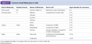
Protein Structure and Function
Classes of Proteins
Proteins serve a wide range of functions in cells, each class specialized for a particular role. - Enzymes: Catalyze biochemical reactions, increasing reaction rates. - Structural proteins: Provide physical support and shape to cells and tissues. - Motility proteins: Enable contraction and movement. - Regulatory proteins: Control and coordinate cellular functions. - Transport proteins: Move substances across cellular membranes. - Signaling proteins: Facilitate communication between cells. - Receptor proteins: Allow cells to respond to environmental stimuli. - Defensive proteins: Protect against disease. - Storage proteins: Serve as reservoirs of amino acids.
Amino Acids: The Monomers of Proteins
Proteins are synthesized from 20 standard amino acids, each with a unique side chain (R group) that determines its properties. - Key Point 1: All amino acids share a common structure: a central α-carbon, an amino group, a carboxyl group, and a variable R group. - Key Point 2: In biological systems, amino acids are almost exclusively found in the L-form. 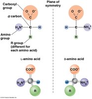
Chemical Structure and Classification of Amino Acids
Amino acids are classified based on the chemical nature of their side chains: nonpolar, polar uncharged, and polar charged. - Key Point 1: The side chain determines the amino acid's role in protein structure and function. - Key Point 2: Glycine is unique among nonpolar amino acids as it does not have separate L and D isomers due to its symmetric structure. 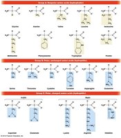
Abbreviations for Amino Acids
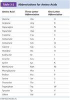
Amino acids are commonly referred to by their three-letter and one-letter abbreviations, which are essential for interpreting protein sequences.
Amino Acid | Three-Letter Abbreviation | One-Letter Abbreviation |
|---|---|---|
Alanine | Ala | A |
Arginine | Arg | R |
Asparagine | Asn | N |
Aspartic acid | Asp | D |
Cysteine | Cys | C |
Glutamine | Gln | Q |
Glutamic acid | Glu | E |
Glycine | Gly | G |
Histidine | His | H |
Isoleucine | Ile | I |
Leucine | Leu | L |
Lysine | Lys | K |
Methionine | Met | M |
Phenylalanine | Phe | F |
Proline | Pro | P |
Serine | Ser | S |
Threonine | Thr | T |
Tryptophan | Trp | W |
Tyrosine | Tyr | Y |
Valine | Val | V |
Peptide Bond Formation
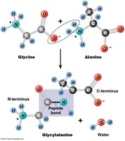
Amino acids are linked together by peptide bonds, formed through dehydration (condensation) reactions. - Key Point 1: The peptide bond is a covalent bond between the carboxyl group of one amino acid and the amino group of another. - Key Point 2: Peptide bond formation releases a molecule of water. 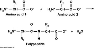
Monomeric and Multimeric Proteins
Proteins may consist of a single polypeptide (monomeric) or multiple polypeptides (multimeric). - Key Point 1: Multimeric proteins are classified by the number of polypeptide subunits: dimers (2), trimers (3), tetramers (4), etc. - Example: Hemoglobin is a tetramer composed of two α and two β subunits. 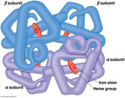
Bonds and Interactions in Protein Folding and Stability
Protein folding and stability depend on both covalent and noncovalent interactions. - Covalent bonds: Peptide bonds and disulfide bonds. - Non-covalent interactions: Hydrogen bonds, ionic bonds, van der Waals forces, and hydrophobic interactions. - Key Point 2: These interactions involve the carboxyl, amino, and R groups of amino acid residues. 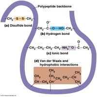
Levels of Protein Structure
Overview of Structural Levels
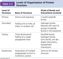
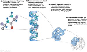
Protein structure is organized into four hierarchical levels: primary, secondary, tertiary, and quaternary.
Level of Structure | Basis of Structure | Kinds of Bonds and Interactions Involved |
|---|---|---|
Primary | Amino acid sequence | Covalent peptide bonds |
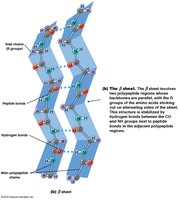
Secondary | Folding into α helix, β sheet, or random coil | Hydrogen bonds between NH and CO groups of peptide bonds in the backbone |
Tertiary | Three-dimensional folding of a single polypeptide chain | Disulfide bonds, hydrogen bonds, ionic bonds, van der Waals interactions, hydrophobic interactions |
Quaternary | Association of multiple polypeptides to form a multimeric protein | Same as for tertiary structure |
Primary Structure
The primary structure is the linear sequence of amino acids in a polypeptide, written from the N-terminus to the C-terminus. - Key Point 1: The sequence is genetically determined by the order of nucleotides in mRNA. - Key Point 2: The order and identity of amino acids direct the formation of higher-order structures. 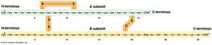
Secondary Structure
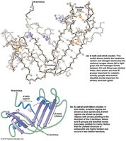
Secondary structure refers to local regions of folding stabilized by hydrogen bonding between backbone NH and CO groups. - Key Point 1: The two major patterns are the α helix and the β sheet. - Key Point 2: Certain amino acids favor specific secondary structures; proline disrupts α helices due to its inability to form hydrogen bonds. 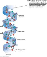
Common Secondary Motifs
Motifs are supersecondary structures composed of a few secondary structure elements, such as β-α-β motifs, hairpin loops, and helix-turn-helix motifs. 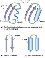
Tertiary Structure
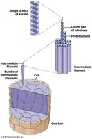
Tertiary structure is the overall three-dimensional shape of a single polypeptide, determined by interactions among R groups. - Key Point 1: Hydrophobic residues avoid water, hydrophilic residues interact with water, and charged residues attract or repel each other. - Key Point 2: Fibrous proteins (e.g., keratin, fibroin) have extensive regions of secondary structure, while globular proteins are compact and folded. 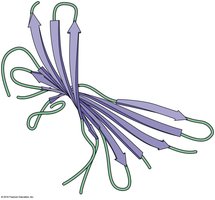
Protein Domains
Domains are distinct structural and functional units within a protein. - Key Point 1: Proteins with similar functions often share common domains. - Key Point 2: Multifunctional proteins may have separate domains for each function. 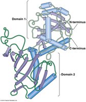
Quaternary Structure
Quaternary structure describes the association of multiple polypeptide chains to form a multimeric protein. - Key Point 1: The same types of bonds and interactions as in tertiary structure maintain quaternary structure. - Example: Hemoglobin's quaternary structure consists of two α and two β subunits.
Protein Folding and Disease
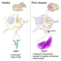
Misfolded proteins can lead to disease, such as prion diseases, which involve abnormal protein aggregation. 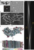

Summary Table: Levels of Protein Structure
Level | Basis | Bonds/Interactions |
|---|---|---|
Primary | Amino acid sequence | Peptide bonds |
Secondary | α helix, β sheet | Hydrogen bonds |
Tertiary | 3D folding | Disulfide, hydrogen, ionic, van der Waals, hydrophobic |
Quaternary | Subunit assembly | Same as tertiary |
Additional info: AlphaFold is a modern AI system for predicting protein tertiary structure, accelerating research in cell biology.
