 뒤로
뒤로Chromosomal Mutations: Variations in Number and Arrangement
스터디 가이드 - 스마트 노트
 자료에 맞춘 맞춤형 노트, 핵심 정의, 예시, 맥락을 확장해 제공합니다.
Chromosomal Mutations
Introduction to Chromosomal Mutations
Chromosomal mutations involve changes at a scale larger than individual genes, affecting the structure or number of entire chromosomes. These modifications can have significant biological consequences, ranging from lethality in animals to phenotypic changes in plants. Because these changes occur at the chromosomal level, they are often heritable and can be passed to future generations.
Types of Chromosomal Modifications: Change in chromosome number, deletions, duplications, and rearrangements.
Biological Impact: Lethal in animals, phenotypic changes in plants, and potential heritability.
Types of Chromosomal Number Variations
Aneuploidy
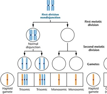
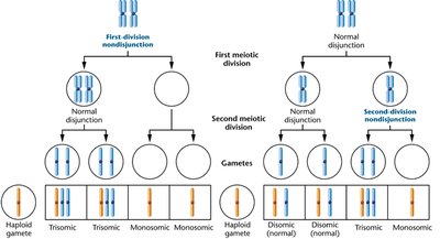
Aneuploidy refers to the presence of an abnormal number of chromosomes in a cell. This can result from the loss or gain of one or more chromosomes, leading to genetic imbalances.
Monosomy: Loss of a single chromosome (2n - 1). Generally not tolerated in animals except for small chromosomes (e.g., X in Turner syndrome).
Trisomy: Gain of an extra chromosome (2n + 1). More tolerated than monosomy, especially for small chromosomes (e.g., trisomy 21 in Down syndrome).
Haploinsufficiency: When a single copy of a gene is insufficient for normal function, leading to phenotypic effects.
Examples of Human Aneuploidy Syndromes
Klinefelter Syndrome (XXY): 47 chromosomes, males with an extra X chromosome.
Turner Syndrome (Monosomy X): 45 chromosomes, females with a single X chromosome.
Trisomy X (XXX): 47 chromosomes, females with an extra X chromosome.
XYY Syndrome: 47 chromosomes, males with an extra Y chromosome.
Down Syndrome (Trisomy 21)
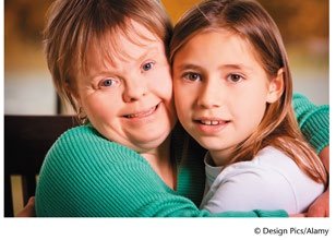
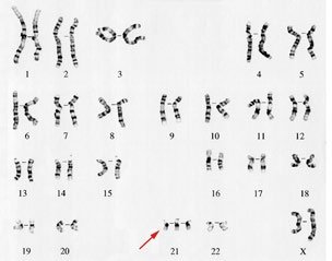
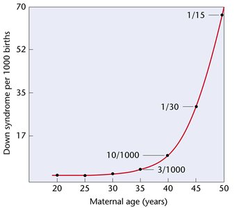
Down syndrome is caused by an extra copy of chromosome 21 (47,21+). The probability of having a child with Down syndrome increases with maternal age. Diagnosis can be performed using karyotyping and various prenatal tests.
Incidence: Approximately 1 in 800 live births in the US.
Detection Methods: Amniocentesis, chorionic villus sampling, non-invasive prenatal genetic diagnosis (NIPGD).
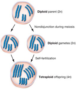
Cause: Usually due to nondisjunction during meiosis.
Other Human Aneuploidy Syndromes
Patau Syndrome (47, 13+): Trisomy of chromosome 13.
Edwards Syndrome (47, 18+): Trisomy of chromosome 18.
Spontaneous Abortions: About 20% of all conceptions abort spontaneously, with 30% of these showing chromosomal imbalance.
Polyploidy
Definition and Types
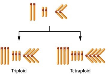
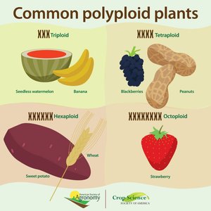
Polyploidy is the condition in which cells have more than two complete sets of chromosomes. It is common in plants and can arise through different mechanisms.
Triploid (3n): Three sets of chromosomes.
Tetraploid (4n): Four sets of chromosomes.
Autopolyploidy: Chromosome duplication within a single species.
Allopolyploidy: Chromosome duplication resulting from hybridization between different species.
Autopolyploidy
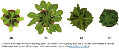
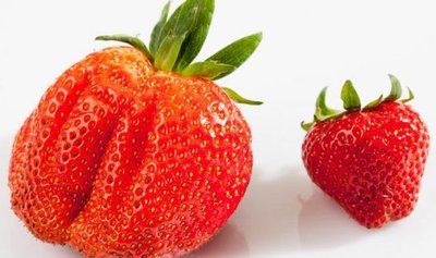
Autopolyploidy results from chromosome duplication within a single species, leading to triploid or tetraploid individuals. These individuals are often larger and may have economic importance, especially in agriculture.
Autotriploids: Can arise from the fusion of a diploid and a haploid gamete.
Autotetraploids: Result from chromosome doubling in a diploid organism.
Phenotypic Effects: Larger flowers and fruits, increased cell size.
Allopolyploidy
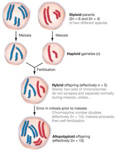
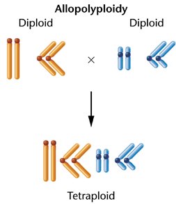
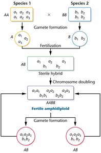
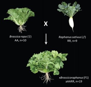
Allopolyploidy occurs when two different species hybridize, and chromosome doubling restores fertility. The resulting organism contains sets of chromosomes from both parent species and is called an amphidiploid if the parental species are known.
Formation: Hybridization followed by chromosome doubling.
Examples: Wheat (Triticum), cotton, and triticale (wheat-rye hybrid).
Endopolyploidy
Endopolyploidy refers to the presence of polyploid cells in otherwise diploid organisms. This phenomenon is observed in certain tissues, such as the liver in humans and salivary glands in some insects.
Examples: Human liver cells (4n, 8n, 16n), water strider salivary glands (up to 2048n).
Possible Function: May increase gene expression in specific tissues.
Chromosomal Rearrangements
Deletions
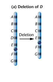
Deletions involve the loss of a chromosome segment. They can be terminal (end of chromosome) or intercalary (internal segment). Deletions can unmask recessive alleles and cause genetic disorders.
Terminal Deletion: Loss of a segment from the end of a chromosome.
Intercalary Deletion: Loss of an internal segment.
Synapsis: Can still occur, but may form a deletion loop during meiosis.
Example: Cri du Chat Syndrome
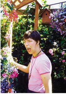
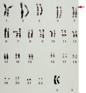
Cri du chat syndrome is caused by a deletion of part of the short arm of chromosome 5 (46, 5p-). It is characterized by malformations, intellectual disability, and a distinctive cry due to abnormal larynx development.
Duplications
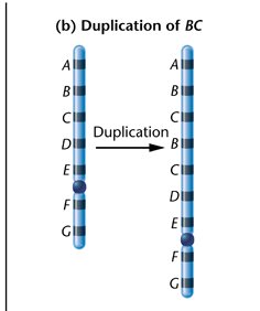
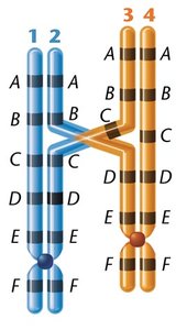
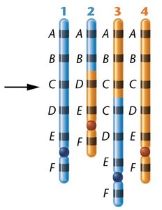
Duplications are chromosomal mutations where a segment of DNA is copied and inserted into the chromosome. They can arise from unequal crossing over or replication errors and may result in compensation loops during meiosis.
Gene Redundancy: Multiple copies of genes, such as rRNA genes, support increased protein synthesis.
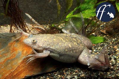
Gene Amplification: Selective replication of certain genes, e.g., rDNA in oocytes of Xenopus laevis.
Gene Duplication in Evolution
Gene duplication provides raw material for evolutionary innovation. Duplicated genes can acquire new functions (neofunctionalization) or divide the original function (subfunctionalization).
Examples: Trypsin and chymotrypsin (digestive enzymes), hemoglobin and myoglobin (oxygen transport).
Gene Families and Copy Number Variations (CNVs)
Gene families are groups of related genes with similar functions, often located near each other on the chromosome. Copy number variations (CNVs) are differences in the number of copies of particular genes among individuals, which can influence disease susceptibility and phenotypic diversity.
CNVs: Can affect 5-10% of the human genome and are linked to diseases such as cancer, autism, and cardiovascular disease.
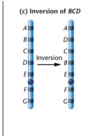
Chromosomal Inversions
Types and Effects
Inversions occur when a chromosome segment is reversed end to end. They do not involve the addition or loss of genetic material but can disrupt gene function and affect meiosis.
Paracentric Inversion: Does not include the centromere.
Pericentric Inversion: Includes the centromere.
Inversion Loops: Formed during meiosis in inversion heterozygotes, leading to abnormal gametes.
Chromosomal Translocations
Types and Mechanisms
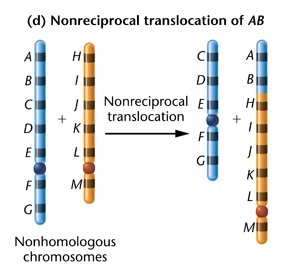
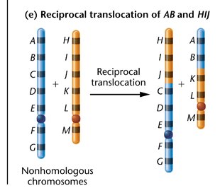
Translocations involve the movement of a chromosomal segment to a non-homologous chromosome. They can be reciprocal (exchange of segments) or nonreciprocal (one-way transfer).
Reciprocal Translocation: Exchange of segments between two non-homologous chromosomes.
Robertsonian Translocation: Fusion of two acrocentric chromosomes, often leading to familial Down syndrome.
Familial Down Syndrome
Familial Down syndrome is caused by a Robertsonian translocation involving chromosomes 14 and 21. One parent is a carrier with a normal phenotype but can pass the translocation to offspring, resulting in Down syndrome.
Summary Table: Types of Chromosomal Mutations
Type | Description | Example |
|---|---|---|
Aneuploidy | Abnormal number of chromosomes | Down syndrome (trisomy 21) |
Polyploidy | More than two sets of chromosomes | Wheat (hexaploid), strawberry (octoploid) |
Deletion | Loss of chromosome segment | Cri du chat syndrome |
Duplication | Repeat of chromosome segment | Bar mutation in Drosophila |
Inversion | Reversal of chromosome segment | Paracentric/pericentric inversions |
Translocation | Segment moves to non-homologous chromosome | Familial Down syndrome |
