 뒤로
뒤로Visualizing Cells: Microscopy Techniques and Molecular Tools in Cell Biology
스터디 가이드 - 스마트 노트
 자료에 맞춘 맞춤형 노트, 핵심 정의, 예시, 맥락을 확장해 제공합니다.
Visualizing Cells
Introduction to Cell Visualization
Understanding the structure and function of cells is fundamental to genetics and cell biology. The ability to visualize cells and their components has advanced significantly due to the development of various microscopy techniques and molecular labeling methods. These tools allow researchers to study cellular architecture, molecular composition, and dynamic processes within living cells.
Microscopy Techniques
Scale and Resolution in Cell Biology
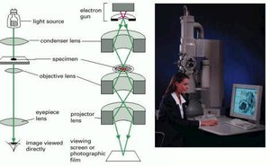
Microscopy enables the observation of structures ranging from whole cells to individual atoms. The resolution of a microscope is determined by the wavelength of the radiation used and the numerical aperture of the lens system.
Resolution Limit: Light microscopes can resolve details as small as 0.2 μm, while electron microscopes can achieve resolutions down to 0.1 nm.
Scale Progression: Magnification increases allow visualization from macroscopic structures (e.g., thumb) to microscopic and molecular levels (e.g., ribosomes, atoms).
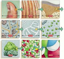
Types of Microscopes
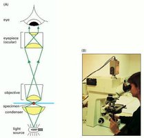
Light Microscope: Uses visible light to illuminate specimens. Essential for observing living cells and tissues.
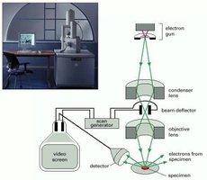
Electron Microscope: Uses electron beams for much higher resolution, allowing visualization of subcellular structures and macromolecules.
Numerical Aperture is a key parameter affecting resolution; higher numerical aperture allows finer detail to be resolved.
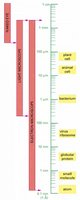
Specimen Preparation and Staining
Specimen preparation is crucial for microscopy. Cells are often fixed, embedded, sectioned, and stained to enhance contrast and reveal specific structures.
Fixation: Immobilizes and preserves cellular structures.
Sectioning: Thin slices are cut using a microtome for high-resolution observation.
Staining: Dyes and stains highlight specific cell components, such as nuclei or mitochondria.
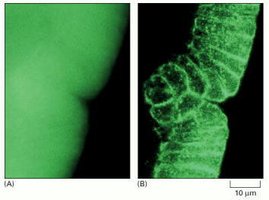
Optical Microscopy: Contrast and Imaging
Different optical techniques are used to visualize living and fixed cells:
Bright-field Microscopy: Simple transmission of light through the specimen.
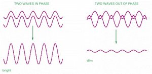
Phase-Contrast Microscopy: Exploits differences in refractive index to visualize unstained living cells.
Differential-Interference-Contrast (DIC) Microscopy: Enhances contrast using interference effects.
Dark-field Microscopy: Illuminates specimens from the side, making them appear bright against a dark background.
Fluorescence Microscopy
Fluorescence microscopy uses fluorescent dyes or proteins to label specific molecules within cells. The optical system includes filters and mirrors to select excitation and emission wavelengths.
Fluorescent Dyes: Absorb light at one wavelength and emit at another, allowing detection of specific molecules.
Common Dyes: DAPI (DNA), GFP (protein), Cy3, Cy5, Alexa dyes.
Immunocytochemistry and Antibody Labeling
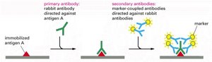
Antibodies are used to detect specific proteins or antigens in cells. Indirect immunocytochemistry amplifies the signal by using secondary antibodies coupled to markers.
Primary Antibody: Binds to the target antigen.
Secondary Antibody: Recognizes the primary antibody and is coupled to a detectable marker (fluorescent dye, enzyme, gold particle).
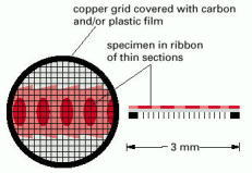
Image Processing and Three-Dimensional Reconstruction
Electronic imaging and computational methods enhance microscopy images and allow three-dimensional reconstructions.
Image Deconvolution: Removes out-of-focus blur to produce crisp optical sections.
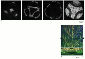
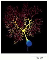
Confocal Microscopy: Uses pinpoint laser illumination and pinholes to exclude out-of-focus light, enabling optical sectioning and 3D reconstruction.
Electron Microscopy
Transmission Electron Microscopy (TEM)
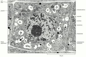
TEM provides high-resolution images of cellular ultrastructure. Specimens are fixed, embedded, sectioned, and stained with heavy metals for contrast.
Resolution: Down to 0.1 nm, revealing organelles and macromolecular complexes.
Preparation: Requires dehydration, embedding, and ultrathin sectioning.
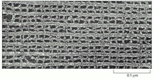
Immunogold Electron Microscopy
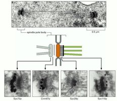
Antibodies coupled to electron-dense gold particles are used to localize specific macromolecules in TEM images.
Three-Dimensional Reconstruction from Serial Sections
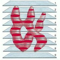
Serial sectioning and reconstruction techniques reveal the true three-dimensional shape of cellular structures.
Scanning Electron Microscopy (SEM)
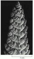
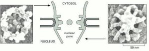
SEM provides detailed images of the surface structure of cells and tissues, with great depth of field and three-dimensional appearance.
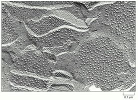
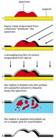
Metal Shadowing and Freeze-Fracture Techniques
These methods allow high-resolution surface views and visualization of membrane interiors.
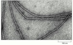
Negative Staining and Cryoelectron Microscopy
Negative staining and cryoelectron microscopy are used to visualize macromolecules and molecular complexes at high resolution.
Combining Multiple Images and Views
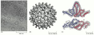
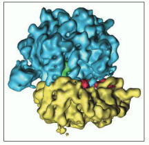
EM tomography combines images from different directions to reconstruct three-dimensional structures of viruses and ribosomes.
Visualizing Molecules in Living Cells
Fluorescent Indicators and Dynamic Imaging
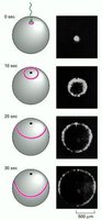
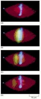
Fluorescent indicators are used to measure ion concentrations and follow dynamic processes in living cells.
Aequorin: A luminescent protein used to monitor Ca2+ concentrations.
Fura-2: A fluorescent indicator for Ca2+ imaging.
Methods for Introducing Molecules into Cells
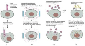
Various methods are used to introduce membrane-impermeant molecules into cells, including microinjection, electroporation, vesicle fusion, and gene delivery.
Caged Molecules and Photoactivation
Caged molecules are inactive precursors that can be activated by light, allowing precise spatial and temporal control of molecular activity in cells.
GFP Tagging and Protein Dynamics
Green fluorescent protein (GFP) and its variants are used to tag proteins genetically, enabling visualization of protein localization and dynamics in living cells and organisms.
Radioisotope Labeling and Autoradiography
Radioisotopes in Cell Biology
Radioisotopes are used to trace the fate of molecules in cells and organisms. Pulse-chase experiments and autoradiography reveal the synthesis, localization, and turnover of biomolecules.
Common Radioisotopes: 32P, 131I, 35S, 14C, 45Ca, 3H
Pulse-Chase: Short labeling followed by nonradioactive chase allows tracking of molecular movement and transformation.
Summary Table: Microscopy Methods and Their Applications
Microscopy Method | Resolution | Application |
|---|---|---|
Light Microscopy | 0.2 μm | Living cells, tissues, basic cell structure |
Fluorescence Microscopy | 0.2 μm | Specific molecules, protein localization |
Confocal Microscopy | 0.2 μm | Optical sectioning, 3D reconstruction |
Transmission Electron Microscopy (TEM) | 0.1 nm | Ultrastructure, organelles, macromolecules |
Scanning Electron Microscopy (SEM) | 10 nm | Surface structure, 3D appearance |
Immunogold Electron Microscopy | 0.1 nm | Localization of specific macromolecules |
Conclusion
Modern microscopy and molecular labeling techniques are essential for visualizing cells and their components, providing insights into cellular structure, function, and dynamics. These methods underpin much of contemporary genetics and cell biology research.
