Ch. 20 - Recombinant DNA Technology
Klug - Concepts of Genetics 12th Edition
Klug12th EditionConcepts of GeneticsISBN: 9780135564776당신이 사용하는 게 아니라요?교과서 변경
20장, 문제 29b

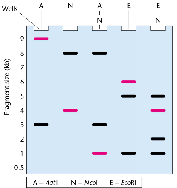
The gel presented here shows the pattern of bands of fragments produced with several restriction enzymes. The enzymes used are identified above the lanes of the gel, and six possible restriction maps are shown in the column to the right.
One of the six restriction maps shown is consistent with the pattern of bands shown in the gel.
The highlighted bands (magenta) in the gel were hybridized with a probe for the gene pep during a Southern blot. Where in the gel is the pep gene located? 
Gel electrophoresis showing DNA fragments from six restriction enzyme digests with magenta bands indicating the pep gene location.

검증된 단계별 안내
1
Step 1: Understand the experimental setup by reviewing how restriction enzymes cut DNA at specific sequences, producing fragments of varying lengths that appear as bands on a gel after electrophoresis.
Step 2: Examine the gel lanes corresponding to each restriction enzyme and note the sizes and number of bands produced, paying special attention to the magenta-highlighted bands that hybridized with the pep gene probe during the Southern blot.
Step 3: Compare the observed band patterns for each enzyme with the six possible restriction maps provided, focusing on which map's predicted fragment sizes and arrangement match the gel's band pattern, especially the bands that hybridize with the pep probe.
Step 4: Identify the fragment(s) in the gel where the pep gene is located by matching the hybridized bands to the corresponding fragment(s) in the restriction map that contains the pep gene sequence.
Step 5: Conclude the location of the pep gene on the restriction map by correlating the hybridized band sizes with the fragment sizes in the map, confirming which restriction map is consistent with the gel pattern and the Southern blot results.

비슷한 문제에 대한 검증된 영상 답변:

이 영상 해법은 위 문제에 도움이 된다고 튜터들이 추천한 것입니다.
영상 길이:
32s
도움이 되었나요?

주요 개념

질문에 올바르게 답하기 위해 반드시 이해해야 하는 핵심 개념들은 다음과 같습니다.

Restriction Enzymes and DNA Fragmentation

Restriction enzymes cut DNA at specific sequences, producing fragments of varying lengths. When DNA is digested with different enzymes, the pattern of fragments reflects the locations of their recognition sites. Understanding how these enzymes work is essential to interpret gel band patterns and map DNA.
추천 영상:
가이드 코스
11:59
Steps to DNA Replication

Gel Electrophoresis and Band Patterns

Gel electrophoresis separates DNA fragments by size, with smaller fragments migrating farther through the gel. The resulting band pattern represents the sizes of DNA pieces after enzyme digestion. Comparing band patterns across lanes helps identify which restriction map matches the observed data.
추천 영상:
가이드 코스
11:19
Segmentation Genes

Southern Blotting and DNA Probes

Southern blotting transfers DNA fragments from a gel to a membrane, where a labeled probe hybridizes to a specific gene sequence. The location of the probe signal on the gel indicates which fragment contains the gene of interest, allowing identification of the gene's position within the restriction map.
추천 영상:
가이드 코스
07:40
Methods for Analyzing DNA and RNA
관련 실천
교과서 질문

Most of the techniques (blotting, cloning, PCR, etc.) are dependent on hybridization (annealing) between different populations of nucleic acids. The length of the strands, temperature, and percentage of GC nucleotides weigh considerably on hybridization. Two other components commonly used in hybridization protocols are monovalent ions and formamide. A formula that takes monovalent Na⁺ ions (M[Na⁺]) and formamide concentrations into consideration to compute a Tₘ (temperature of melting) is as follows:

Tₘ=81.5+16.6(log M[Na+])+0.41(%GC)−0.72(%formamide)

For the following concentrations of Na⁺ and formamide, calculate the Tₘ. Assume 45% GC content.

  [Na⁺]  % Formamide

  0.825      20

  0.825      40

  0.165      20

  0.165      40

526
views
교과서 질문

The CRISPR-Cas system has great potential but also raises many ethical issues about its potential applications because, theoretically, it can be used to edit any gene in the genome. What do you think are some of the concerns about the use of CRISPR-Cas on humans? Should CRISPR-Cas applications be limited for use on only certain human genes but not others? Explain your answers.

680
views
교과서 질문

Gene targeting and gene editing are both techniques for removing or modifying a particular gene, each of which can produce the same ultimate goal. What is the main technical difference in how DNA is modified that differs between these approaches?

732
views
교과서 질문

Most of the techniques described in this chapter (blotting, cloning, PCR, etc.) are dependent on hybridization (annealing) between different populations of nucleic acids. The length of the strands, temperature, and percentage of GC nucleotides weigh considerably on hybridization. Two other components commonly used in hybridization protocols are monovalent ions and formamide. A formula that takes monovalent Na⁺ ions (M[Na⁺]) and formamide concentrations into consideration to compute a Tₘ (temperature of melting) is as follows:

Tₘ=81.5+16.6(log M[Na+])+0.41(%GC)−0.72(%formamide)

Given that formamide competes for hydrogen bond locations on nucleic acid bases and monovalent cations are attracted to the negative charges on nucleic acids, explain why the Tₘ varies as described in part (a).

449
views
교과서 질문

A widely used method for calculating the annealing temperature for a primer used in PCR is 5 degrees below the melting temperature, Tₘ(°C), which is computed by the equation 81.5+0.41×(%GC)−(675/N), where %GC is the percentage of GC nucleotides in the oligonucleotide and N is the length of the oligonucleotide. Notice from the formula that both the GC content and the length of the oligonucleotide are variables. Assuming you have the following oligonucleotide as a primer,

5′-TTGAAAATATTTCCCATTGCC-3′

Compute the annealing temperature for PCR. What is the relationship between %GC and? Why? (Note: In reality, this computation provides only a starting point for empirical determination of the most useful annealing temperature.)

1016
views
교과서 질문

The gel presented here shows the pattern of bands of fragments produced with several restriction enzymes. The enzymes used are identified above the lanes of the gel, and six possible restriction maps are shown in the column to the right.

One of the six restriction maps shown is consistent with the pattern of bands shown in the gel.

From your analysis of the pattern of bands on the gel, select the correct restriction map and explain your reasoning.

593
views