 뒤로
뒤로Peripheral Vascular and Lymphatic Systems: Structure, Function, and Clinical Assessment
스터디 가이드 - 스마트 노트
 자료에 맞춘 맞춤형 노트, 핵심 정의, 예시, 맥락을 확장해 제공합니다.
Peripheral Vascular System
Overview
The peripheral vascular system consists of the arteries, veins, and lymphatic vessels that transport blood and lymph throughout the body. This system is essential for delivering oxygen and nutrients to tissues and removing waste products. Disorders of the peripheral vascular system can lead to significant morbidity, including pain, tissue damage, and impaired organ function.
Arteries
Structure and Function
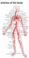
Arteries are high-pressure vessels that carry oxygenated blood away from the heart to the tissues.
They have thick, muscular walls to withstand and regulate blood pressure.
Major palpable arteries include: temporal, carotid, brachial, ulnar, radial, femoral, popliteal, posterior tibial, and dorsalis pedis.
Veins
Structure and Function
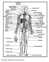
Veins are low-pressure vessels that return deoxygenated blood and waste products from the tissues to the heart.
They have thinner walls and larger lumens than arteries, and often contain valves to prevent backflow.
Veins lie closer to the skin surface and are classified as superficial or deep.
Major veins include: jugular, subclavian, radial, medial, basilic, cephalic (arms); femoral, popliteal, great saphenous, small saphenous (legs).
Great Vessels
Major Arteries and Veins Connected to the Heart
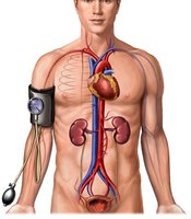
Arteries: The left ventricle pumps blood into the aorta; the right ventricle pumps blood into the pulmonary artery.
Veins: The superior and inferior vena cava return blood to the right atrium; the pulmonary veins return oxygenated blood to the left atrium.
Lymphatic System
Structure and Function
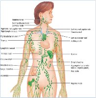
The lymphatic system is a network of vessels and nodes that retrieves excess fluid from tissues, filters it, and returns it to the bloodstream. It also plays a key role in immune defense.
Right lymphatic duct drains the right upper body; thoracic duct drains the rest of the body.
Major lymphatic organs: spleen, tonsils, thymus.
Lymph Nodes
Small, oval clumps of lymphatic tissue located along lymphatic vessels.
Filter lymph fluid before it returns to the bloodstream.
Swollen, tender nodes indicate local inflammation or infection.
Superficial node groups: cervical, axillary, epitrochlear, inguinal.
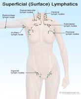
Developmental Competence
Age-Related Differences
Infants and Children: Lymph nodes are larger and more prominent.
Pregnant Females: Vasodilation occurs, increasing risk of edema and varicosities.
Older Adults: Arteriosclerosis (hardening of arteries) is common, increasing risk of vascular disease.
Clinical Assessment of the Peripheral Vascular System
Subjective Data Collection
Ask about changes in skin color, temperature, or texture.
Assess for pain in the legs, especially if it awakens the patient from sleep (may indicate arterial disease).
Inquire about varicose veins, edema, or changes in sexual activity (in males, impotence may indicate vascular insufficiency).
Ask about lymph node enlargement, duration, and associated symptoms.
Objective Data Collection
Inspect and palpate upper and lower extremities for skin changes, hair distribution, lesions, ulcers, edema, atrophy, symmetry, and venous patterns.
Assess pulses: radial, brachial, femoral, popliteal, posterior tibial, dorsalis pedis.
Grade pulses: 3+ (bounding), 2+ (normal), 1+ (weak), 0 (absent).
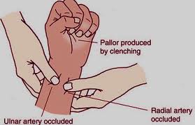
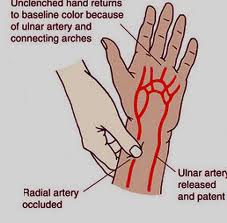
Modified Allen’s Test
This test evaluates the patency of the radial and ulnar arteries in the hand.
First, occlude both arteries and have the patient clench their fist, producing pallor.
Next, release one artery and observe for return of color, indicating adequate blood flow.
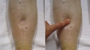
Pitting Edema
Graded on a scale from 1+ (slight indentation) to 4+ (indentation lasts several minutes).
Arterial Deficit Assessment
With the patient supine, elevate legs to drain venous blood, then observe skin color.
Have the patient sit with legs dangling and observe for return of color.
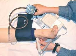
Ankle-Brachial Index (ABI)
The ABI is used to assess the severity of peripheral arterial disease (PAD).
Calculated as:
Normal ABI: 1.0–1.4; ABI ≤ 0.90 indicates PAD.
Peripheral Vascular Disease (PVD)
Leg Ulcers
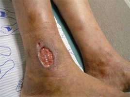
Ischemic (arterial) ulcers: Occur on toes, heels, or ankles; typically do not bleed and are associated with shiny skin and thick nails.
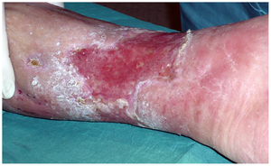
Venous (stasis) ulcers: Occur at the medial malleolus; are red, swollen, and may weep.
Pulse Variations
Weak (thready) pulse: Indicates decreased cardiac output or peripheral arterial disease.
Bounding pulse: May be seen in hyperkinetic states.
Pulsus bigeminus: Every other beat is premature.
Pulse alternans: Alternating strong and weak beats, often seen in heart failure.
Nursing Interventions for PVD
Assess edema and peripheral pulses every 4 hours.
Evaluate pain and provide meticulous foot care daily.
Maintain extremity warmth and provide patient education.
PVD in Arms
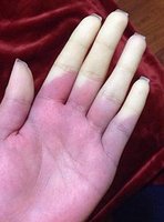
Raynaud’s phenomenon: Vasospasm of arteries causing color changes in fingers/toes (white, blue, red).
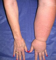
Lymphedema: Swelling due to lymphatic obstruction, often after lymph node removal or damage.
PVD in Legs (cont.)
Varicose veins: Dilated, tortuous superficial veins due to valve incompetence.
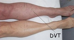
Deep vein thrombosis (DVT): Formation of a blood clot in a deep vein, causing swelling, pain, and risk of embolism.
Arterial vs. Venous Insufficiency
Feature | Arterial (PAD) | Venous (PVD) |
|---|---|---|
Pain | Intermittent claudication, deep muscle pain, throbbing | Aching, tiredness, chronic pain worse at end of day |
Skin | Cool, pale, shiny, thick ridged nails | Red, warm, swollen, thickened, brown discoloration |
Ulcers | On toes, heels, ankles; do not bleed | At medial malleolus; weeping, jagged edges |
Other | Dependent rubor, motor & sensory loss | Edema, varicosities |
Health Promotion
Check feet daily for injuries or changes.
Maintain blood flow by regular movement and exercise.
Wear comfortable, well-fitting shoes.
Keep skin soft and smooth to prevent breakdown.
Sample Clinical Question
Question: A nurse is assessing a client who has chronic peripheral arterial disease (PAD). Which of the following findings should the nurse expect?
Pallor on elevation of the limbs, and rubor when the limbs are dependent.
