 뒤로
뒤로Ch.2 Body Structure: Levels of Organization, Body Planes, and Cavities
스터디 가이드 - 스마트 노트
 자료에 맞춘 맞춤형 노트, 핵심 정의, 예시, 맥락을 확장해 제공합니다.
Levels of Body Organization
Overview of Body Organization
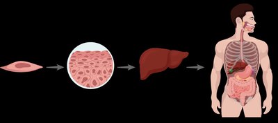
The human body is organized into several hierarchical levels, each building upon the previous one to form the complete organism. Understanding these levels is fundamental to medical terminology and anatomy.
Cellular Level: The smallest functional unit of all living organisms. Cells contain a cell membrane, cytoplasm, and a nucleus.
Tissue Level: Groups of similar cells working together to perform related functions.
Organ Level: Structures composed of two or more types of tissues working together for a specific function.
Organ System Level: Groups of organs that coordinate to perform complex functions.
Cell Structure and Function
Cells are the basic building blocks of the body. Each cell contains:
Cell Membrane: Provides protection, enables communication, and regulates the exchange of substances in and out of the cell.
Cytoplasm: Gel-like substance that surrounds internal structures and supports cellular processes.
Nucleus: Contains genetic material (DNA) and acts as the control center for cell reproduction, metabolism, and growth.
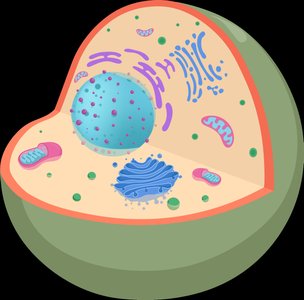
Example: The nucleus contains genetic material and acts as the control center of the cell.
Tissue Types
Tissues are groups of similar cells that work together to perform specific functions. There are four main types of tissues:
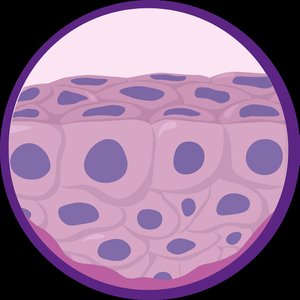
Epithelial Tissue: Covers surfaces and lines cavities; functions include protection, absorption, secretion, and filtration.
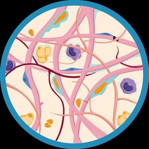
Connective Tissue: Found in bones, cartilage, tendons, blood, and fat; provides structure, support, and protection.
Muscular Tissue: Includes skeletal (attached to bone), smooth (internal organs), and cardiac (heart) muscle; contracts to create movement.
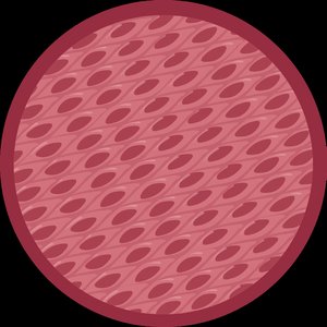
Nervous Tissue: Found in the brain, spinal cord, and nerves; detects stimuli and transmits signals.
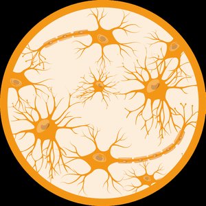
Example: Connective tissue supports and binds other tissues like bones and tendons.
Organs and Organ Systems
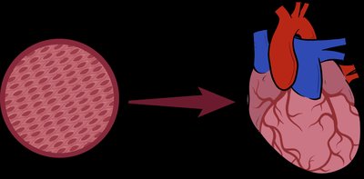
Organs are structures composed of two or more types of tissues working together for a specific function. Organ systems are groups of organs that coordinate to perform complex functions.
Integumentary System: Skin, hair, nails; protection and temperature regulation.
Skeletal System: Bones, joints, cartilage; structure and protection.
Muscular System: Muscles; movement.
Nervous System: Brain, spinal cord, nerves; communication and control.
Cardiovascular System: Heart, arteries, veins; transport of substances and waste removal.
Blood System: Blood; transport of substances.
Lymphatic System: Lymph nodes, spleen, tonsils; immune defense.
Endocrine System: Pituitary gland, adrenal gland; communication and control.
Respiratory System: Lungs, trachea; gas exchange.
Digestive System: Stomach, intestines, liver; ingestion, digestion, absorption.
Urinary System: Kidneys, bladder; filtration and removal of waste.
Reproductive System: Ovaries, testes; reproduction.
Body Planes and Directions
Body Planes
Body planes are imaginary surfaces used to divide the body into sections for anatomical reference and description. The standard anatomical position is standing upright, feet together, arms at sides, and palms facing forward.
Frontal (Coronal) Plane: Divides the body into anterior (front) and posterior (back) portions.
Sagittal Plane: Divides the body into right and left portions.
Transverse (Horizontal) Plane: Divides the body into superior (upper) and inferior (lower) portions.
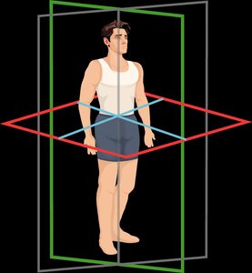
Example: The transverse plane separates the urinary bladder and cranium.
Body Directions and Positions
Directional terms describe the location of body parts relative to each other and the body's position. These are essential for accurate communication in medical settings.
Medial: Toward the midline of the body.
Lateral: Away from the midline.
Proximal: Closer to the point of attachment or origin.
Distal: Farther from the point of attachment or origin.
Superior (Cranial): Toward the head or upper part of a structure.
Inferior (Caudal): Away from the head or toward the lower part of a structure.
Anterior (Ventral): Toward the front of the body.
Posterior (Dorsal): Toward the back of the body.
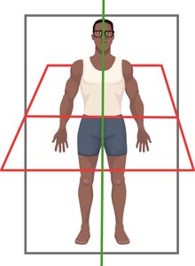
Example: The foot is inferior to the heart, and the hips are superior to the heart.
Body Positions
Body positions are used to describe a patient's posture during examinations and surgical procedures.
Supine: Lying face up.
Prone: Lying face down.
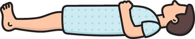

Example: Spinal fusion surgery is typically performed with the patient in the prone position.
Body Cavities
Overview of Body Cavities
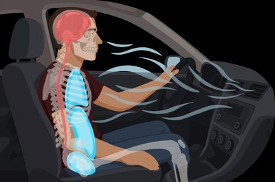
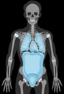
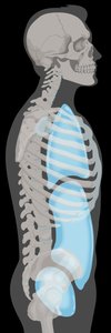
Body cavities are hollow spaces that house internal organs. They are classified into two main groups: ventral (anterior) and dorsal (posterior) cavities. Cavities protect, support, and separate organs.
Ventral Body Cavities
Thoracic Cavity: Contains the lungs (pleural cavities) and heart (pericardial cavity), separated from the abdominal cavity by the diaphragm.
Abdominal Cavity: Contains the stomach, intestines, and other digestive organs.
Pelvic Cavity: Contains the urinary bladder and reproductive organs.
Dorsal Body Cavities
Cranial Cavity: Space inside the skull, contains the brain.
Spinal Cavity: Contains the spinal cord and spinal fluid.
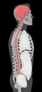

Example: The organs of the central nervous system are in the dorsal cavities.
Abdominopelvic Regions and Quadrants
Regions and Quadrants
The abdominopelvic cavity is divided into nine regions and four quadrants for anatomical reference, diagnosis, and surgical purposes.
Regions: Right/Left Hypochondriac, Epigastric, Right/Left Lumbar, Umbilical, Right/Left Inguinal (Iliac), Hypogastric.
Quadrants: Right Upper (RUQ), Left Upper (LUQ), Right Lower (RLQ), Left Lower (LLQ).
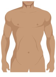
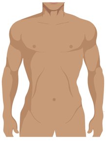
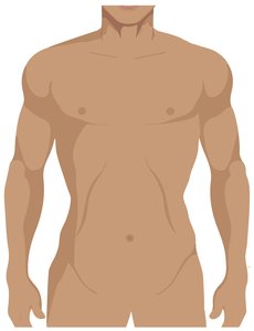
Example: The urinary bladder is located in the hypogastric region.
Key Organs in Quadrants
RUQ: Liver, right kidney, gallbladder
LUQ: Stomach, spleen, left kidney
RLQ: Appendix, right ovary & fallopian tube, right ureter
LLQ: Left ovary & fallopian tube, left ureter
Example: The gallbladder is present only in the right upper quadrant (RUQ).
Summary Table: Levels of Organization
Level | Definition | Example |
|---|---|---|
Cell | Smallest functional unit | Muscle cell |
Tissue | Group of similar cells | Muscle tissue |
Organ | Group of tissues | Heart |
Organ System | Group of organs | Cardiovascular system |
