 뒤로
뒤로Medical Terminology: Nervous System and Mental Health – Study Notes
스터디 가이드 - 스마트 노트
 자료에 맞춘 맞춤형 노트, 핵심 정의, 예시, 맥락을 확장해 제공합니다.
Overview of the Nervous System
Introduction to the Nervous System
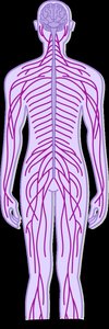
The nervous system is an intricate communication network that coordinates the activities of the body. It consists of the brain, spinal cord, and nerves, and is responsible for receiving, processing, and transmitting information throughout the body. The nervous system is divided into two main parts: the Central Nervous System (CNS) and the Peripheral Nervous System (PNS).
Central Nervous System (CNS): Composed of the brain and spinal cord. It receives, processes, and interprets sensory information and controls consciousness and voluntary actions.
Peripheral Nervous System (PNS): Consists of all nerves outside the CNS. It transmits information between the CNS and the rest of the body, including sensory and motor functions.
Nervous Tissue
Types of Nervous Tissue
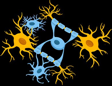
Nervous tissue makes up most of the nervous system and consists of two main types of cells: neurons and neuroglia.
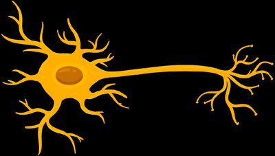
Neurons: The structural and functional units of the nervous system. They conduct electrical signals and respond to stimuli. Each neuron has dendrites (branched extensions that receive signals) and an axon (a long extension that transmits signals).
Neuroglia (Glial Cells): Provide support and protection to neurons. They do not conduct electrical signals but produce myelin, which forms the myelin sheath around axons for insulation.
Central Nervous System (CNS)
Composition of the CNS
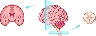
The CNS is composed of two types of matter: white matter and gray matter.
White Matter: Contains myelinated axons, optimized for rapid signal transmission.
Gray Matter: Contains unmyelinated neuron cell bodies and dendrites. In the brain, the outer layer (cortex) is gray matter, while the inner layer is white matter. In the spinal cord, the arrangement is reversed.
Major Regions of the Brain
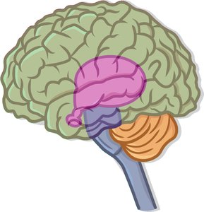
The brain coordinates most body activities and is the center of thought, memory, judgment, and emotion. It is divided into four major regions:
Cerebrum: The largest part, responsible for sensory and motor activity, divided into four lobes.
Diencephalon: Central part of the brain, acts as a relay for sensory and motor information (includes thalamus, epithalamus, hypothalamus).
Brainstem: Controls vital involuntary functions (midbrain, pons, medulla oblongata).
Cerebellum: Located posteriorly, integrates sensory perception with motor activity.
Lobes of the Cerebrum
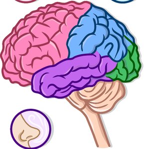
The cerebrum consists of four lobes, each named after the cranial bones that overlie them and each with specialized functions:
Frontal Lobe: Controls voluntary movement, planning, decision making, consciousness, and personality.
Parietal Lobe: Processes sensation, temperature, touch, and spatial perception.
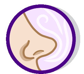
Temporal Lobe: Responsible for hearing, smell, and memory.
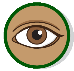
Occipital Lobe: Responsible for vision and visual association.


Spinal Cord Structure and Function
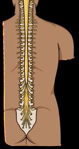
The spinal cord is a column of nervous tissue with a core of gray matter surrounded by white matter. It contains tracts, which are bundles of nerve fibers that interconnect parts of the CNS:
Ascending Tracts: Carry sensory information up to the brain.
Descending Tracts: Carry motor information down from the brain.

Peripheral Nervous System (PNS)
Introduction to the PNS
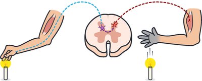
The PNS includes all nervous tissue outside the CNS. Nerves are bundles of axons that can be classified by the direction of information flow:
Afferent (Sensory) Nerves: Transmit sensory information to the CNS.
Efferent (Motor) Nerves: Transmit motor information from the CNS to effectors (muscles and glands).
Mnemonic: SAME – Sensory Afferent, Motor Efferent.
Subdivisions of the PNS
The PNS is divided into two functional subdivisions:
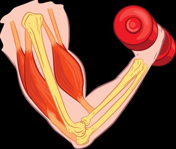
Somatic Nervous System (SNS): Controls voluntary movements by serving skeletal muscles.
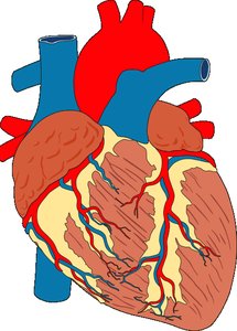
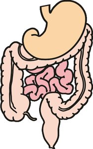
Autonomic Nervous System (ANS): Controls involuntary functions by serving smooth muscle, cardiac muscle, and glands.
Branches of the Autonomic Nervous System (ANS)
The ANS is further divided into two branches with generally opposite actions:
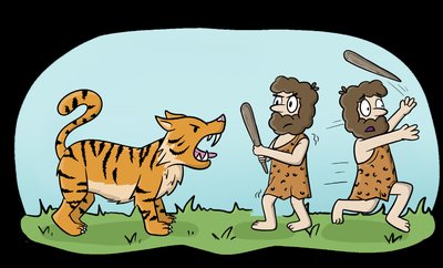
Sympathetic Branch: Activates during stress or threat ("fight-or-flight"), increases heart rate, dilates airways, raises blood pressure, and inhibits digestion.
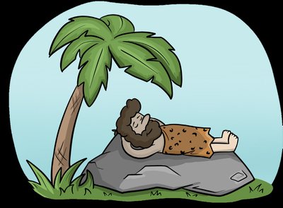
Parasympathetic Branch: Activates during relaxation ("rest-and-digest"), decreases heart rate, constricts airways, lowers blood pressure, and stimulates digestion.
Nervous System: Word Building
Combining Forms for Structures
Medical terminology uses combining forms to describe structures of the nervous system. Below is a table summarizing key combining forms:
Combining Form | Definition | Memory Tool |
|---|---|---|
cephal/o | head | En/cephal/o: inside head |
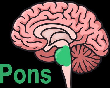
pont/o | pons | — |
dur/o | dura mater, tough membrane | Durable hard hat |
lamin/o | lamina, plate | Lamination |
poli/o | gray matter | Pale gray |
gli/o | glue, glial cells | Gleeful gluey cell |
gangli/o | ganglion, swelling | Gang of knots |
astr/o | star | astr/o/nomy |
radic/o, radicul/o | nerve root | Radish root |


Combining Forms for Functions and Pathology
Additional combining forms describe functions and pathology of the nervous system:
Combining Form | Definition | Memory Tool |
|---|---|---|
esthesi/o | sensation, feeling | an/esthesi/a |
alges/o | pain | an/alges/ic |
olfact/o | smell | Smelly ol-factory |
son/o, ech/o | sound | son/ar |
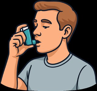
hal/o | to breathe | in/hal/er |
somn/o | sleep | in/somn/ia |
ambul/o | to walk | Walking hospital |
clon/o | rapid contracting and relaxing | Twitchy Clown |
topic/o | place, area | Topic/al cream |
isch/o | to hold back, block | Scheduled time block |
thromb/o | clot | Tied down thromb/us |
embol/o | embolus (plug) | Escaped embol/us |
Nervous System Disorders
Common Disorders and Their Definitions
Below is a table summarizing key nervous system disorders, their definitions, and memory tools:
Medical Term | Definition | Memory Tool |
|---|---|---|
Bell’s Palsy | Sudden and temporary unilateral facial paralysis | Bells Pausing |
Cerebral Palsy | Brain damage to motor control center during childbirth | Cerebrum Pausing |
Huntington’s Chorea | Genetic disorder with jerky, involuntary movements | Hunt for the Choreographer |
Guillain-Barré Syndrome | Acute, autoimmune disorder destroying myelin sheath | Growing weakness |
Multiple Sclerosis | Chronic, autoimmune disorder destroying myelin sheath | My Sheaths are Scrambled |
Parkinson’s Disease | Progressive neurological disorder with dopamine cell loss | No Dope in the PARK |
Alzheimer’s Disease | Chronic, degenerative disorder with neuron death and dementia | Forgetful Old Timer |
Reye Syndrome | Triggered by viral infection and aspirin use in children | All Sick Pediatrics Invite Risk If Not careful |
Sciatica | Pain, numbness, tingling down the sciatic nerve | Sci-fi Attack |
Spina Bifida | Birth defect with incomplete closure of spinal column | Spine Bulge |
Akathisia | Inability to stay still | Antsy Kat can’t sit still |
Medical Procedures and Terms
Key Procedures and Clinical Terms
Medical Term | Definition | Memory Tool |
|---|---|---|
Babinski’s Sign | Stroking the foot causes big toe to move upward | BabinSKI: Big Toe Skiing up |
Deep Tendon Reflex | Muscle contraction from a hammer hitting a tendon | Knee-Deep Reflex |
Romberg Test | Measures physical balance | Rom-balance |
Spasticity | Unusually high muscle tone | Surge of Tone |
Tremor | Involuntary, repetitive movement | Trembling |
Aura | Sensations before seizures or migraines | An Unusual Reaction Activates Senses |
Syncope | Fainting due to low cranial blood pressure | SINK on me |
Burr Holes | Openings drilled in skull to relieve pressure | Bring Urgent Relief Rapidly |
Hydrocephalus Shunt | Tube to drain excess fluid from brain | hydro- = water, cephal/o = head, shunt = drain |
Positron Emission Tomography (PET Scan) | Imaging technique using radioactive sugar | Pets Eat Tons of Sugar |
Neurotransmitters and Pharmacology
Key Neurotransmitters and Drugs
Medical Term | Definition | Memory Tool |
|---|---|---|
Acetylcholine (ACH) | Neurotransmitter for muscle contraction and memory | ACHing Muscles and Memory |
Endorphins | Neurotransmitters that reduce pain and enhance well-being | EndorPhins: End Pain |
Dopamine | Neurotransmitter for movement, pleasure, and motivation | It’s Dope feeling good |
Anticonvulsants | Drugs that prevent or control seizures | Anti-commotion |
Levodopa | Treats dopamine deficiency in the brain | LEVO-tating DOPe levels |
