 뒤로
뒤로Medical Terminology: Special Senses – The Eye and Ear
스터디 가이드 - 스마트 노트
 자료에 맞춘 맞춤형 노트, 핵심 정의, 예시, 맥락을 확장해 제공합니다.
Special Senses: The Eye and Ear
Overview of the Eye
The eye is a specialized organ responsible for vision, containing sensory receptors that convert light into nerve impulses. In addition to the eyeball, several external structures protect and support visual function.
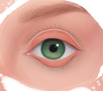
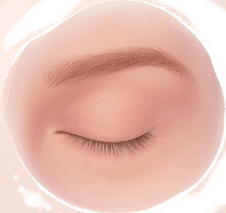
Eyelids & Eyelashes: Serve as physical barriers, protecting the eye from foreign particles and injury. Sebaceous glands in the eyelids lubricate the eye surface.
Conjunctiva: A protective mucous membrane covering the underside of the eyelids and the anterior surface of the eyeball.
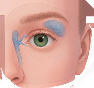
Lacrimal Apparatus: Includes lacrimal glands and ducts, responsible for producing tears that wash and lubricate the eye.
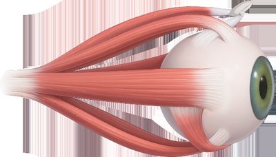
Eye Muscles: Six skeletal muscles attach the eyeball to the skull, controlling eye movement.

Structure of the Eyeball
The eyeball is a globe-shaped, fluid-filled organ composed of three main layers, each with distinct functions:
Outer Layer: Includes the cornea (transparent, bends light rays) and sclera (white, tough coating for protection and muscle attachment).
Middle Layer (Uvea): Contains the iris (colored part, controls light entry), ciliary body (holds lens, produces aqueous humor), and choroid (pigmented, prevents internal light reflection).
Inner Layer: The retina contains photoreceptor cells—rods (dim light, gray tones) and cones (bright light, color vision). The optic nerve transmits visual information to the brain.
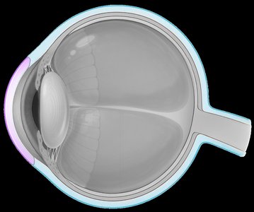
Pathway of Light Through the Eye
Light enters the eye through the cornea, passes through the pupil (center of the iris), is focused by the lens, and reaches the retina, where photoreceptors convert it to nerve impulses sent to the brain via the optic nerve.
Word Building: Visual System
Medical terminology for the visual system uses combining forms, prefixes, and suffixes to describe structures, functions, and diseases.
Combining Form | Definition |
|---|---|
ocul/o, ophthalm/o | eye |
opt/o, optic/o | vision, eye |
blephar/o | eyelid |
ir/o, irid/o | iris |
cor/o | pupil |
cycl/o | ciliary body |
phac/o, lent/o | lens |
papill/o | optic disk |
dacry/o, lacrim/o | tear, lacrimal apparatus |
chromat/o | color |
Common Suffixes and Prefixes
Suffix/Prefix | Definition |
|---|---|
-opia, -opsia | vision condition |
-tropia | eye misalignment |
-pexy | surgical fixation |
my- | to shut (as in myopia) |
Visual System Pathology
Several medical terms describe disorders of the visual system:
Cataract: Clouding of the lens, leading to impaired vision.
Chalazion: Firm, non-tender cyst on the eyelid due to blocked gland.
Hordeolum (Stye): Painful, infected cyst on the eyelid.
Strabismus: Misaligned eyes, often due to muscle weakness.
Myopia: Nearsightedness; difficulty seeing distant objects.
Retinitis Pigmentosa: Progressive degeneration of the retina, leading to blindness.


Diagnostic and Surgical Procedures
Snellen Test: Measures visual acuity using a chart of letters at 20 feet.
Keratometry: Measures corneal curvature.
Gonioscopy: Examines the anterior chamber angle of the eye.
Exophthalmometry: Measures protrusion of the eyeball.
Enucleation: Surgical removal of the eyeball.

Glaucoma: Pathogenesis and Treatment
Glaucoma is characterized by increased intraocular pressure due to impaired outflow or overproduction of aqueous humor, leading to optic nerve damage and vision loss.
Pathogenesis: Blocked drainage ducts cause aqueous humor to accumulate, raising intraocular pressure.
Treatment: Medications include prostaglandin analogues (increase outflow), beta blockers (decrease production), alpha adrenergic antagonists, carbonic anhydrase inhibitors, and cholinergic agonists (increase pupil constriction to enhance outflow).

Overview of the Ear
Ear Anatomy and Function
The ear is responsible for hearing and equilibrium, divided into three anatomical regions:
External Ear: Includes the auricle (funnels sound waves) and auditory canal (transmits sound inward). Cerumen (earwax) protects the canal.
Middle Ear: Air-filled cavity containing three ossicles (malleus, incus, stapes) that amplify vibrations from the tympanic membrane (eardrum) to the inner ear.
Inner Ear: Fluid-filled cavity (labyrinth) containing the cochlea (converts vibrations to nerve impulses) and vestibular apparatus (equilibrium).
Word Building: Auditory System
Combining Form | Definition |
|---|---|
acous/o, audi/o, audit/o | hearing |
aur/o, auricul/o | ear |
myring/o, tympan/o | eardrum, tympanic membrane |
Suffix | Definition |
|---|---|
-cusis, -acusus | hearing |
-otia | ear condition |
-ory | pertaining to |
Auditory Pathology
Tinnitus: Perception of internal noise (ringing) in one or both ears.
Vertigo: Sensation of spinning or loss of balance.
Ménière’s Disease: Excess inner ear fluid causing tinnitus, vertigo, and hearing loss.
Diagnostic and Surgical Procedures: Auditory System
Decibel (dB): Unit measuring sound loudness.
Hertz (Hz): Unit measuring sound frequency (pitch).
Rinne Test: Assesses conduction of sound in one ear at a time.
Weber Test: Assesses conduction of sound in both ears simultaneously.
Fenestration: Surgical creation of an opening in the inner ear to treat otosclerosis.
Brainstem Auditory-Evoked Responses (BAERs): Neurological test for brain response to auditory stimuli.
Electronystagmography: Diagnoses ear and equilibrium disorders by measuring eye movements.
