 뒤로
뒤로Skeletal System: Medical Terminology Study Guide
스터디 가이드 - 스마트 노트
 자료에 맞춘 맞춤형 노트, 핵심 정의, 예시, 맥락을 확장해 제공합니다.
Ch.4 Skeletal System
Overview of the Skeletal System
The skeletal system is a fundamental component of the human body, providing structure, protection, movement, production, and storage. It consists of bones, cartilage, ligaments, and joints, each serving specific functions essential for health and mobility.
Support: The skeleton forms the framework of the body, maintaining its shape and posture.
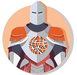
Protection: Bones protect vital organs such as the brain, heart, and lungs from injury.
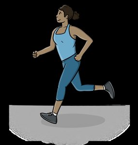
Movement: Bones act as levers and joints as fulcrums, enabling movement when muscles contract.
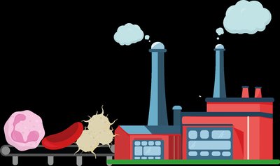
Production: The skeletal system produces blood cells in the bone marrow, including red blood cells, white blood cells, and platelets.
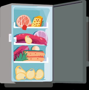
Storage: Bones store minerals such as calcium and phosphorus, which are essential for various bodily functions.
Functions of Specific Bones
Mandible (Lower jawbone): Supports chewing and speech (Support, Movement).
Ribs: Protect the lungs and heart (Protection).
Spinal column: Provides structural support and flexibility (Support, Movement).
Femur (Thigh bone): Bears body weight and enables movement (Support, Movement).
Classification of Bones
Types of Bones
Bones are classified based on their shape and function. Most bones in the human body are long bones, but other types exist to serve specialized roles.
Long Bones: Elongated shape; found in arms and legs (e.g., femur).
Short Bones: Cube-shaped; found in wrists and ankles (e.g., carpals).
Flat Bones: Thin and broad; found in the shoulder blade (scapula), ribs, and sternum.
Irregular Bones: Complex shapes; found in vertebrae and facial bones.
Sesamoid Bones: Develop within tendons; found in the kneecap (patella).
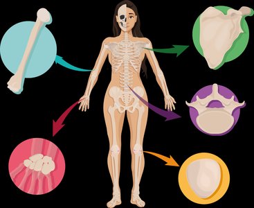
Examples of Bone Classification
Femur: Long bone
Sternum: Flat bone
Carpals: Short bone
Patella: Sesamoid bone
Mandible: Irregular bone
Parts of the Skeleton
Skeletal Organization
The human skeleton contains 206 bones and is organized into two main parts: the axial skeleton and the appendicular skeleton. Each part has distinct functions and anatomical structures.
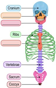
Axial Skeleton: Includes the skull, vertebral column, and thoracic cage. Provides support and protection for the brain, spinal cord, and thoracic organs.
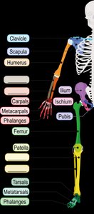
Appendicular Skeleton: Includes the limbs and the pectoral and pelvic girdles. Facilitates movement and interaction with the environment.
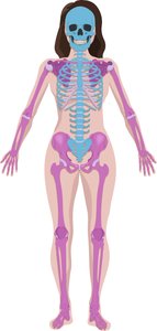
Major Bones of the Axial Skeleton
Skull: Cranium, maxilla (upper jaw), mandible (lower jaw)
Spinal Column: Vertebrae, sacrum, coccyx (tailbone)
Thoracic Cage: Ribs, sternum (breastbone)
Major Bones of the Appendicular Skeleton
Pectoral Girdle: Clavicle (collarbone), scapula (shoulder blade)
Upper Limbs: Humerus (upper arm), radius and ulna (forearm), carpals, metacarpals, phalanges (hand)
Pelvic Girdle: Ilium, ischium, pubis
Lower Limbs: Femur (thigh), patella (kneecap), tibia (shinbone), fibula (lower leg), tarsals, metatarsals, phalanges (foot)
Bone Markings
Surface Features of Bones
Bones have various markings that serve as attachment points for muscles, passageways for nerves and blood vessels, and articulation sites for joints.
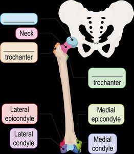
Head: Rounded end of a long bone that helps in joint formation.
Condyle: Smooth, rounded projections at the lower end of a bone.
Trochanter: Large, rough processes for muscle attachment.
Neck: Area between the head and the trochanters.
Bone Depressions
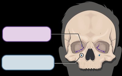
Fossa: Shallow depression at the surface of a bone.
Fissure: Slit-like opening.
Foramen: Smooth, rounded opening.
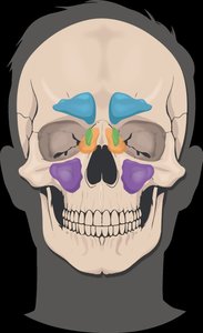
Sinus: Hollow cavity within a bone.
Joints and Movements
Types of Joints
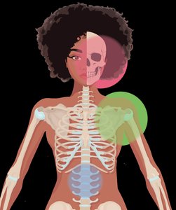
Joints, or articulations, are classified based on their degree of movement:
Synarthrosis: Immovable joint (e.g., skull sutures).
Amphiarthrosis: Slightly movable joint (e.g., intervertebral discs).
Diarthrosis: Freely movable joint (e.g., shoulder, hip).
Joint Movements
Freely movable joints (diarthroses) allow a variety of movements, each described by specific medical terms.
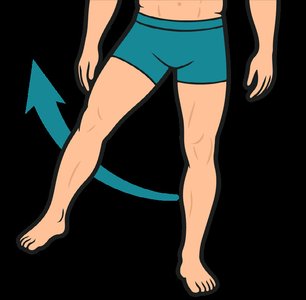
Abduction: Moving a body part away from the midline.
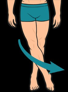
Adduction: Moving a body part toward the midline.
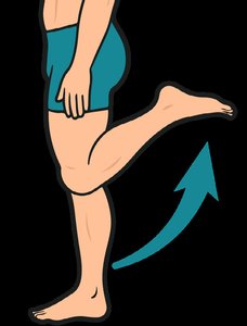
Flexion: Bending a joint, decreasing the angle between bones.
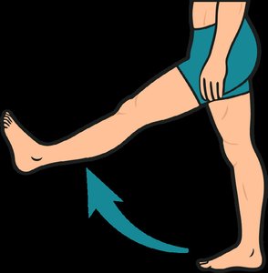
Extension: Straightening a joint, increasing the angle between bones.
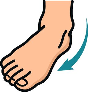
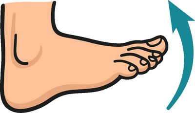
Dorsiflexion: Bending the ankle upward.
Plantar Flexion: Bending the ankle downward toward the sole.
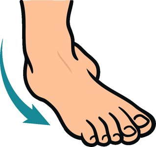
Inversion: Turning the sole of the foot inward.
Eversion: Turning the sole of the foot outward.
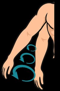
Pronation: Turning the palm or foot downward.
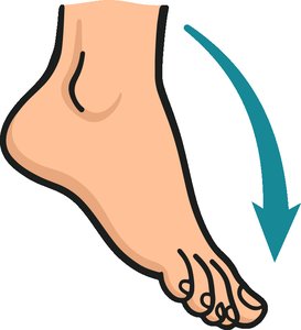
Supination: Turning the palm or foot upward.
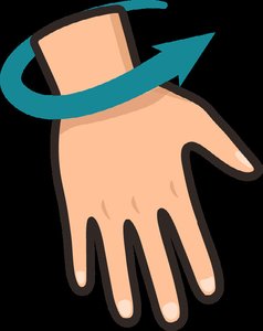
Protraction: Moving a body part forward.
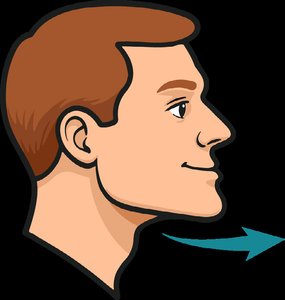
Retraction: Moving a body part backward.
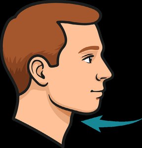
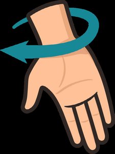
Circumduction: Moving a body part in a circular motion.
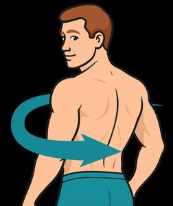
Rotation: Turning a body part around its axis.
Skeletal System: Word Building
Combining Forms
Medical terminology for the skeletal system often uses combining forms based on anatomical names and characteristics.
crani/o: cranium
humer/o: humerus
sacr/o: sacrum
tibi/o: tibia
cervic/o: neck
cost/o: ribs
olecran/o: elbow
condyl/o: knuckle
chir/o: hand
ped/o, pod/o: foot
dactyl/o: finger, toe
articul/o: joint
myel/o: bone marrow, spinal cord
spondyl/o: vertebra
coll/a: glue
syn/o: connect
burs/o: sac
acr/o: extremity
Characteristics and Conditions
ankyl/o: stiff, fused
scoli/o: crooked, curvature
kyph/o: humpback
lord/o: bent backward
orth/o: straight
fixat/o: fastened
rheumat/o: discharge
lith/o: stone
por/o: pore
prosthet/o: artificial part
Suffixes
-clast, -clasia: to break
-listhesis: slipping
-phyte: growth
-poiesis: formation
-lysis: loosen
-oid: resembling
-dynia: pain
-ion: process
-ous: pertaining to
Skeletal System Vocabulary
Pathology and Fractures
Rheumatoid arthritis: Autoimmune disease marked by joint inflammation.
Genu valgum (Knock-knee): Knees touch, ankles apart.
Genu varum (Bowleg): Knees angle out, ankles together.
Gout: Acute arthritis marked by foot joint inflammation.
Rickets: Disease causing weak, soft bones from vitamin D deficiency.
Hallux: Great toe.
Acetabulum: Cup-shaped socket in the pelvis forming the hip joint.
Symphysis: Joint where two bones are connected by fibrocartilage.
Comminuted fracture: Bone broken into three or more pieces.
Compound fracture: Bone breakage through the skin.
Compression fracture: Bone breakage from pressure leading to collapse.
Greenstick fracture: Incomplete bone breakage, typical in children.
Oblique fracture: Bone breakage at an angle.
Transverse fracture: Complete bone breakage straight across.
Medical Procedures
Surgical and Diagnostic Terms
Reduction: Realignment of bone fragments.
Traction: Continuous pulling force to restore alignment.
Spinal Fusion: Permanently joining multiple bones of the spine.
DEXA Scan: Measures bone density using low-dose X-rays.
CT Scan: Computerized scanning system providing cross-sectional and 3D images.
MRI Scan: Body imaging method using radio waves in a magnetic field.
Summary Table: Bone Types and Functions
Bone | Type | Main Function |
|---|---|---|
Femur | Long bone | Support, Movement |
Ribs | Flat bone | Protection |
Vertebrae | Irregular bone | Support |
Patella | Sesamoid bone | Movement |
Carpals | Short bone | Movement |
Summary Table: Types of Joints
Joint Type | Movement | Example |
|---|---|---|
Synarthrosis | Immovable | Skull sutures |
Amphiarthrosis | Slightly movable | Intervertebral discs |
Diarthrosis | Freely movable | Shoulder, hip |
