Skip to main content
Microbiology
나의 코스
배우기
시험 준비
AI 튜터
학습 가이드
교과서적 해결책
플래시카드
탐험해 보세요
앱을 사용해 보세요
나의 코스
배우기
시험 준비
AI 튜터
학습 가이드
교과서적 해결책
플래시카드
탐험해 보세요
앱을 사용해 보세요
뒤로
Light Microscopes that Increase Contrast definitions
카드를 뒤집기 위해 탭할 수 있습니다.
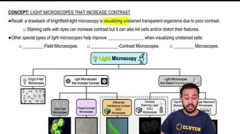
Bright Field Microscope
카드를 뒤집기 위해 탭할 수 있습니다.
👆
Bright Field Microscope
Instrument producing images with a dark specimen on a bright background, often requiring staining for better visibility.
진행 상황 추적
컨트롤 버튼이 '내비게이션' 모드로 변경되었습니다.
1/14
관련 플래시카드
관련 실천
추천 영상
Light Microscopes that Increase Contrast quiz
Light Microscopes that Increase Contrast
15 용어
Light Microscopes that Increase Contrast
9. Microscopes
2 문제점
주제
Nicole
Light Microscopes that Detect Fluorescence
9. Microscopes
5 문제점
주제
Monica
9. Microscopes - Part 1 of 3
6 주제
12 문제점
장
Nicole
9. Microscopes - Part 2 of 3
8 주제
10 문제점
장
Nicole
9. Microscopes - Part 3 of 3
1 주제
3 문제점
장
02:39
Light Microscopes that Increase Contrast
2395
views
44
rank
01:52
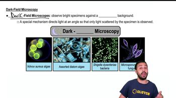
Dark-Field Microscopy
2105
views
28
rank
03:27
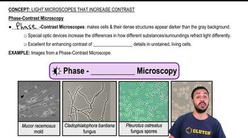
Phase-Contrast Microscopy
2293
views
35
rank
이 집합의 용어 (14)
하이드의 정의
Bright Field Microscope
Instrument producing images with a dark specimen on a bright background, often requiring staining for better visibility.
Contrast
Degree of visual difference between a specimen and its background, crucial for distinguishing cell features.
Staining
Process using dyes to color cells, enhancing visibility but potentially killing or distorting specimens.
Dark Field Microscope
Instrument creating images with a bright specimen on a dark background by detecting only scattered light.
Phase Contrast Microscope
Device using special optics to amplify light refraction differences, revealing internal details in unstained living cells.
Differential Interference Contrast Microscope
Instrument generating highly detailed, three-dimensional images with added color contrast using advanced optics.
Optic Devices
Complex components within microscopes that manipulate light to enhance image contrast and reveal fine details.
Background
Visual field surrounding the specimen, which can be manipulated to improve specimen visibility in microscopy.
Three-dimensional Image
Visual output providing depth and detail, making specimens appear to extend off the viewing surface.
Unstained Cell
Living specimen observed without dyes, maintaining natural features but often requiring special microscopy for visibility.
Internal Structure
Cellular components and organelles made visible by advanced contrast techniques without the need for staining.
Scattered Light
Illumination redirected by a specimen, used in certain microscopes to enhance the specimen's visibility.
Grayish Background
Neutral field often produced in phase contrast microscopy, making cells and structures appear darker for better contrast.
Live Specimen
Organism or cell observed in its natural, undisturbed state, often requiring non-destructive imaging methods.
 뒤로뒤로
뒤로뒤로
 02:39
02:39