Skip to main content
Microbiology
나의 코스
배우기
시험 준비
AI 튜터
학습 가이드
교과서적 해결책
플래시카드
탐험해 보세요
앱을 사용해 보세요
나의 코스
배우기
시험 준비
AI 튜터
학습 가이드
교과서적 해결책
플래시카드
탐험해 보세요
앱을 사용해 보세요
뒤로
Light Microscopes that Increase Contrast quiz
카드를 뒤집기 위해 탭할 수 있습니다.
What is a major drawback of bright field light microscopy when visualizing unstained transparent organisms?
카드를 뒤집기 위해 탭할 수 있습니다.
👆
What is a major drawback of bright field light microscopy when visualizing unstained transparent organisms?
Bright field light microscopy has poor contrast, making it difficult to visualize unstained transparent organisms.
진행 상황 추적
컨트롤 버튼이 '내비게이션' 모드로 변경되었습니다.
1/15
관련 플래시카드
관련 실천
추천 영상
Light Microscopes that Increase Contrast definitions
Light Microscopes that Increase Contrast
14 용어
Light Microscopes that Increase Contrast
9. Microscopes
2 문제점
주제
Nicole
Light Microscopes that Detect Fluorescence
9. Microscopes
5 문제점
주제
Monica
9. Microscopes - Part 1 of 3
6 주제
12 문제점
장
Nicole
9. Microscopes - Part 2 of 3
8 주제
10 문제점
장
Nicole
9. Microscopes - Part 3 of 3
1 주제
3 문제점
장
02:39
Light Microscopes that Increase Contrast
2394
views
44
rank
01:52
Dark-Field Microscopy
2103
views
28
rank
03:27
Phase-Contrast Microscopy
2291
views
35
rank
이 집합의 용어 (15)
하이드의 정의
What is a major drawback of bright field light microscopy when visualizing unstained transparent organisms?
Bright field light microscopy has poor contrast, making it difficult to visualize unstained transparent organisms.
Why might scientists avoid staining cells with dyes when using light microscopes?
Staining dyes can kill cells or distort their features, which may not be desirable for certain studies.
Name three types of light microscopes that increase contrast for unstained cells.
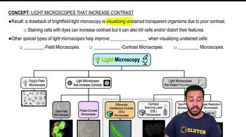
Dark field, phase contrast, and differential interference contrast (DIC) microscopes increase contrast for unstained cells.
How does dark field microscopy differ from bright field microscopy in terms of specimen appearance?
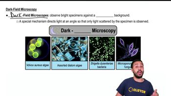
Dark field microscopy shows a bright specimen against a dark background, while bright field shows a dark specimen against a light background.
What mechanism does dark field microscopy use to enhance contrast?
Dark field microscopy directs light at an angle so only light scattered by the specimen is observed, enhancing contrast.
What is the main visual feature of images produced by dark field microscopes?
Specimens appear bright against a dark background, making them easy to distinguish.
What defines phase contrast microscopes?
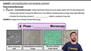
Phase contrast microscopes use complex optical devices to amplify differences in light refraction between substances.
How do cells and their structures appear in phase contrast microscopy images?
Cells and their structures appear darker than the typically grayish background.
What is a key advantage of phase contrast microscopy over bright field microscopy?
Phase contrast microscopy allows visualization of internal details in unstained living cells without requiring staining.
Do all phase contrast microscopes produce images with gray backgrounds?
No, some variations of phase contrast microscopes do not produce gray backgrounds.
What is the main function of the special optic devices in phase contrast microscopes?
They increase the differences in how substances refract light, enhancing contrast.
What type of images do DIC microscopes produce?
DIC microscopes produce highly detailed, three-dimensional images of live specimens.
How are the special optic devices in DIC microscopes similar to those in phase contrast microscopes?
Both use special optic devices to enhance contrast, but DIC adds contrasting colors and 3D imaging.
What is a distinguishing feature of DIC microscopy compared to phase contrast microscopy?
DIC microscopy creates three-dimensional images and adds contrasting colors.
Why are light microscopes that increase contrast important for studying live, unstained cells?
They allow visualization of cell features without killing or altering the cells, providing clearer and more accurate images.
 뒤로뒤로
뒤로뒤로
 02:39
02:39