 뒤로
뒤로Cell Structure and Function: Prokaryotic and Eukaryotic Cells
스터디 가이드 - 스마트 노트
 자료에 맞춘 맞춤형 노트, 핵심 정의, 예시, 맥락을 확장해 제공합니다.
Cell Structure and Function
Overview of Prokaryotic and Eukaryotic Cells
Cells are the fundamental units of life, classified as either prokaryotic or eukaryotic. This distinction is based on structural and functional differences, which are foundational to understanding microbiology.
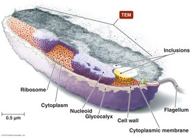
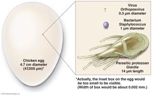
Prokaryotic cells include members of the domains Bacteria and Archaea. They lack a nucleus and membrane-bound organelles, and are typically smaller (1.0 µm or less in diameter).
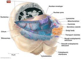
Eukaryotic cells belong to the domain Eukarya and possess a true nucleus and various internal membrane-bound organelles. They are generally larger (10–100 µm in diameter) and more complex.
Cell Theory, developed by Schwann and Schleiden, states that all living organisms are composed of cells, which are the basic units of life and arise from pre-existing cells.
Processes of Life
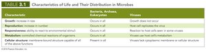
All living organisms share four essential processes: growth, reproduction, responsiveness, and metabolism. These processes distinguish living cells from non-living matter and viruses.
Growth: Increase in size.
Reproduction: Production of new organisms, either asexually or sexually.
Responsiveness: Ability to respond to environmental changes.
Metabolism: All chemical reactions that provide energy and materials for growth, reproduction, and responsiveness.
Characteristic | Bacteria, Archaea, Eukaryotes | Viruses |
|---|---|---|
Growth | Occurs in all | Does not occur |
Reproduction | Occurs in all | Host cell replicates the virus |
Responsiveness | Occurs in all | Reaction to host cells seen in some viruses |
Metabolism | Occurs in all | Viruses lack metabolism |
Cellular structure | Present in all | Viruses lack cytoplasmic membrane or cellular structure |
Bacterial Cell Structures
External Structures of Bacterial Cells
Bacteria possess several external structures that contribute to their survival, pathogenicity, and ability to interact with their environment.
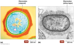
Glycocalyx: A gelatinous, sticky substance surrounding the cell, composed of polysaccharides, polypeptides, or both. It exists as either a capsule (organized, firmly attached) or a slime layer (loose, water-soluble). Capsules can help bacteria evade the host immune system, while slime layers aid in surface attachment and biofilm formation.
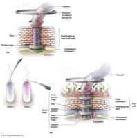
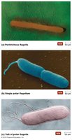
Flagella: Long, whip-like structures responsible for motility. They consist of a filament, hook, and basal body. The arrangement and structure of flagella can be used to classify bacteria (e.g., peritrichous, polar, endoflagella).
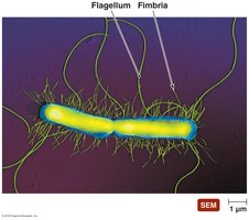
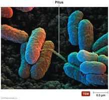
Fimbriae and Pili: Fimbriae are short, bristle-like projections that help bacteria adhere to surfaces and each other, playing a key role in biofilm formation. Pili (conjugation pili) are longer and involved in DNA transfer between cells (conjugation).
Bacterial Cell Walls
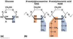
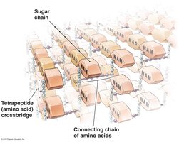
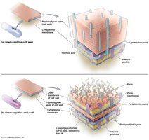
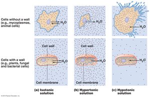
The cell wall provides structural support, shape, and protection from osmotic forces. It is primarily composed of peptidoglycan, a mesh-like polymer of sugars (NAG and NAM) and amino acids.
Gram-positive cell walls: Thick peptidoglycan layer, teichoic acids, sometimes mycolic acid (acid-fast bacteria).
Gram-negative cell walls: Thin peptidoglycan layer, outer membrane with lipopolysaccharide (LPS), periplasmic space.
Bacteria without cell walls: Mycoplasmas and L-form bacteria lack cell walls but have sterol-containing membranes.
Bacterial Cytoplasmic Membranes
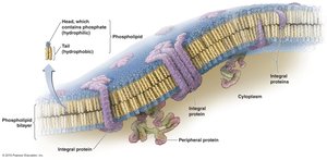
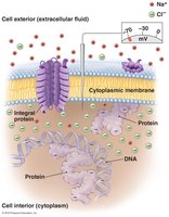
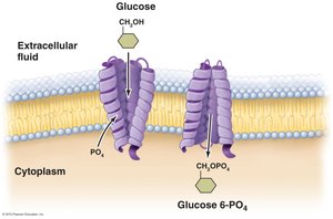
The cytoplasmic membrane is a phospholipid bilayer with embedded proteins, described by the fluid mosaic model. It controls the movement of substances into and out of the cell and maintains chemical and electrical gradients.
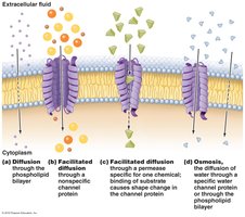
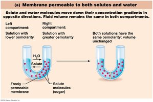
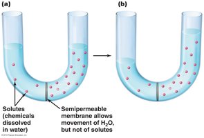
Passive transport: Includes diffusion, facilitated diffusion, and osmosis. No energy is required.
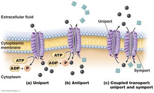
Active transport: Requires energy (ATP or ion gradients) and includes uniport, antiport, symport, and group translocation mechanisms.
Cytoplasm of Bacteria
The cytoplasm is the gelatinous interior of the cell, containing cytosol, inclusions, and non-membranous organelles.
Cytosol: The liquid portion, site of many metabolic reactions, and location of the nucleoid (bacterial chromosome).
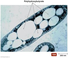
Inclusions: Reserve deposits of nutrients or other substances (e.g., polyhydroxybutyrate, gas vesicles, magnetite crystals).
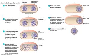
Endospores: Highly resistant, dormant structures formed by some bacteria (e.g., Bacillus, Clostridium) for survival under adverse conditions.
Ribosomes: Sites of protein synthesis (70S in prokaryotes).
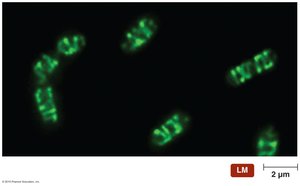
Cytoskeleton: Protein fibers that maintain cell shape and aid in division and movement.
Archaeal Cell Structures
External and Internal Structures of Archaea
Archaea share some structural similarities with bacteria but also possess unique features that allow them to thrive in extreme environments.
Glycocalyces, flagella, and fimbriae are present, but archaeal flagella are thinner, not hollow, and powered by ATP.
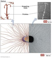
Hami: Unique, grappling hook-like structures used for attachment.
Cell walls: Composed of polysaccharides or proteins, lacking peptidoglycan.
Cytoplasmic membranes: Contain ether-linked lipids, providing stability in extreme conditions.
Cytoplasm: Contains 70S ribosomes, circular DNA, and a fibrous cytoskeleton. Ribosomal proteins and genetic code are more similar to eukaryotes than bacteria.
Eukaryotic Cell Structures
External Structures and Cell Walls
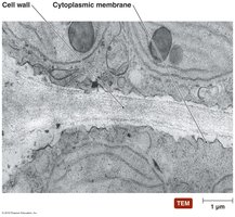
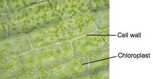
Eukaryotic cells may have a glycocalyx (in animal and protozoan cells) or a cell wall (in fungi, algae, and plants). The glycocalyx aids in cell recognition and protection, while cell walls provide structural support and prevent osmotic lysis.
Plant cell walls: Composed of cellulose.
Fungal cell walls: Composed of cellulose, chitin, and/or glucomannan.
Algal cell walls: Composed of various polysaccharides and chemicals (e.g., agar, carrageenan, silicates).
Eukaryotic Cytoplasmic Membranes
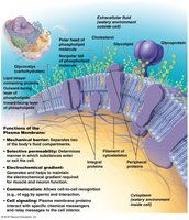
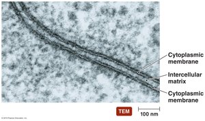
The eukaryotic cytoplasmic membrane is a fluid mosaic of phospholipids, proteins, glycolipids, and cholesterol. It controls the movement of substances and participates in cell signaling and attachment.
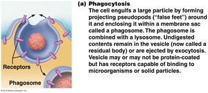
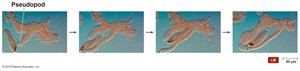
Vesicular transport: Includes endocytosis (phagocytosis, pinocytosis, receptor-mediated) and exocytosis, allowing bulk movement of materials.
Motility Structures: Flagella and Cilia
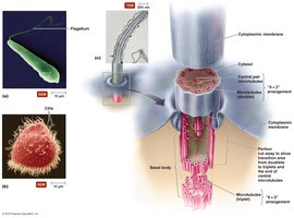
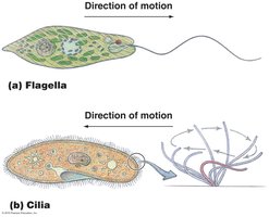
Eukaryotic flagella and cilia are internal extensions of the cytoskeleton, composed of microtubules arranged in a "9 + 2" pattern. Flagella undulate for movement, while cilia beat rhythmically to move the cell or substances past its surface.
Non-membranous Organelles
Ribosomes: Larger (80S) than prokaryotic ribosomes, composed of 40S and 60S subunits.
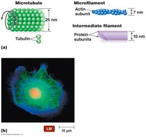
Cytoskeleton: Includes microfilaments (actin), intermediate filaments, and microtubules (tubulin), providing shape, support, and movement.
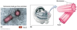
Centrioles and centrosomes: Involved in cell division and formation of cilia and flagella.
Membranous Organelles
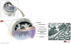
Nucleus: Contains most of the cell's DNA, surrounded by a double membrane with nuclear pores. The nucleolus is the site of ribosome assembly.
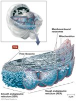
Endoplasmic reticulum (ER): Rough ER synthesizes proteins; smooth ER is involved in lipid metabolism and detoxification.
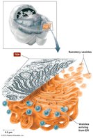
Golgi apparatus: Modifies, packages, and sorts proteins and lipids for secretion or use within the cell.
Lysosomes and peroxisomes: Contain enzymes for digestion and detoxification.
Vacuoles: Storage and maintenance of cellular homeostasis, especially in plants and protists.
Mitochondria: Sites of cellular respiration and ATP production; contain their own DNA and 70S ribosomes.
Chloroplasts: Sites of photosynthesis in plants and algae; also contain their own DNA and 70S ribosomes.
Endosymbiotic Theory
The endosymbiotic theory proposes that mitochondria and chloroplasts originated as free-living prokaryotes that were engulfed by ancestral eukaryotic cells. Evidence includes the presence of double membranes, circular DNA, and 70S ribosomes in these organelles.
Summary Table: Comparison of Prokaryotic and Eukaryotic Cells
Feature | Prokaryotes | Eukaryotes |
|---|---|---|
Nucleus | Absent | Present |
Membrane-bound organelles | Absent | Present |
Cell wall composition | Peptidoglycan (bacteria), proteins/polysaccharides (archaea) | Cellulose (plants), chitin (fungi), various (algae) |
Ribosome size | 70S | 80S (cytoplasm), 70S (mitochondria/chloroplasts) |
DNA | Circular, single chromosome | Linear, multiple chromosomes |
Size | 1–10 µm | 10–100 µm |
