 뒤로
뒤로Cell Structure and Function: Prokaryotic and Eukaryotic Cells
스터디 가이드 - 스마트 노트
 자료에 맞춘 맞춤형 노트, 핵심 정의, 예시, 맥락을 확장해 제공합니다.
Cell Structure and Function
Overview of Prokaryotic and Eukaryotic Cells
Cells are the fundamental units of life and are classified into two main types: prokaryotic and eukaryotic cells. These classifications are based on structural and functional differences, particularly the presence or absence of a nucleus and membrane-bound organelles.
Prokaryotic cells lack a true nucleus and membrane-bound organelles. They include members of the domains Bacteria and Archaea.
Eukaryotic cells possess a nucleus and various membrane-bound organelles. They include organisms in the domain Eukarya (e.g., plants, animals, fungi, and protists).
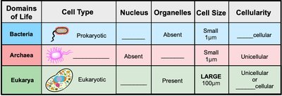
Key Differences Between Prokaryotic and Eukaryotic Cells
Nucleus: Prokaryotes lack a nucleus; eukaryotes have a nucleus.
Organelles: Prokaryotes do not have membrane-bound organelles; eukaryotes do.
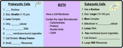
Cell Size: Prokaryotic cells are generally smaller (1–10 μm) than eukaryotic cells (10–100 μm).
Cellularity: Prokaryotes are almost always unicellular; eukaryotes can be unicellular or multicellular.
DNA Structure: Prokaryotic DNA is typically circular and found in a region called the nucleoid; eukaryotic DNA is linear and housed within the nucleus.
Ribosomes: Prokaryotes have 70S ribosomes; eukaryotes have 80S ribosomes.
Cell Division: Prokaryotes divide by binary fission; eukaryotes divide by mitosis and cytokinesis.
Structure of Bacterial and Eukaryotic Cells
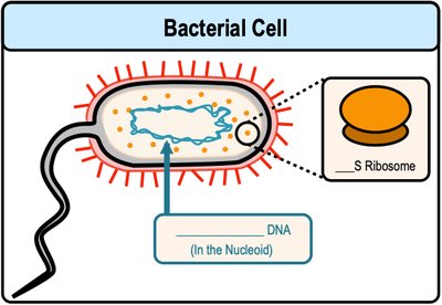
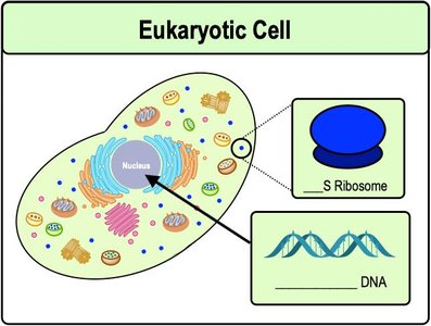
Bacterial cells are prokaryotic and have a simple structure, while eukaryotic cells are more complex.
Bacterial Cell: Contains circular DNA in the nucleoid, small 70S ribosomes, and lacks membrane-bound organelles.
Eukaryotic Cell: Contains linear DNA within a nucleus, large 80S ribosomes, and various organelles such as mitochondria, endoplasmic reticulum, and Golgi apparatus.
Shared Features of All Cells
All cells have a cell membrane composed of a phospholipid bilayer.
All cells contain genetic material (DNA), ribosomes for protein synthesis, and cytoplasm.
Prokaryotic Cell Structures
Structures Outside and Inside the Cell
Prokaryotic cells possess various structures that contribute to their survival and function.
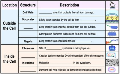
Location | Structure | Description |
|---|---|---|
Outside the Cell | Cell Wall | Rigid layer that protects the cell from damage. |
Outside the Cell | Glycocalyx | Sticky layer secreted by the cell to form capsules or slime layers. |
Outside the Cell | Flagella | Long protein filaments used for cell motility. |
Outside the Cell | Pili/Fimbriae | Protein filaments that extend from the cell surface for attachment or DNA transfer. |
Inside the Cell | Ribosomes | Site of protein synthesis in the cytoplasm. |
Inside the Cell | Inclusions | Molecular aggregates for storage in the cytoplasm. |
Inside the Cell | Endospores | Dormant cell type resistant to damaging conditions. |
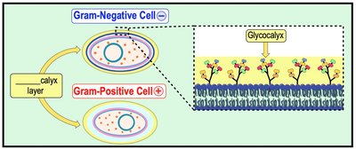
The Glycocalyx: Capsules and Slime Layers
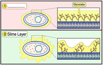
The glycocalyx is a gelatinous, sticky substance that surrounds the outside of some bacterial cells. It is composed mainly of polysaccharides and sometimes polypeptides.
Capsule: Highly organized and firmly attached to the cell wall; provides protection against desiccation and immune defenses.
Slime Layer: Loosely organized and easily removed; aids in attachment to surfaces and formation of biofilms.
Biofilms
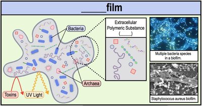
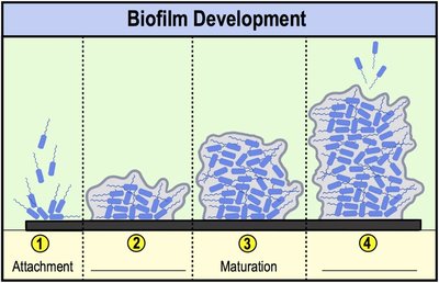
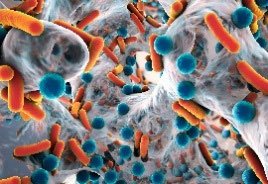
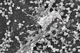
Biofilms are structured communities of microorganisms encased in a self-produced matrix of extracellular polymeric substances (EPS) and attached to a surface. Biofilms provide protection from environmental threats such as toxins, UV light, and antibiotics.
Biofilms are important in medical and environmental contexts due to their resistance to antimicrobial agents.
Development occurs in stages: attachment, colonization, maturation, and dispersal.
Quorum Sensing
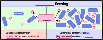
Quorum sensing is a cell-to-cell communication process that enables bacteria within a biofilm to detect and respond to cell population density by producing and sensing signaling molecules called autoinducers. This process regulates gene expression and coordinates group behaviors such as biofilm formation and virulence.
Prokaryotic Motility: Flagella
Flagellar Structure and Function
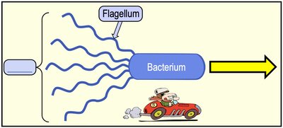
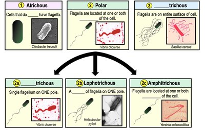
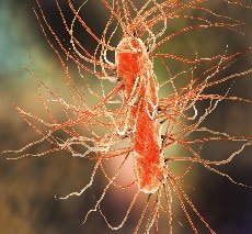
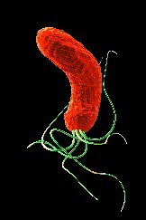
Flagella are long, whip-like appendages that provide motility to many prokaryotic cells. The arrangement and number of flagella can be used to classify bacteria.
Monotrichous: Single flagellum at one pole.
Lophotrichous: Tuft of flagella at one or both poles.
Amphitrichous: One flagellum at each pole.
Peritrichous: Flagella distributed over the entire cell surface.
Atrichous: No flagella.

Flagellar Structure
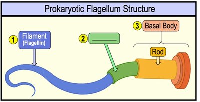
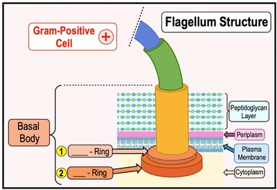
A typical prokaryotic flagellum consists of three main parts:
Filament: Composed of the protein flagellin; forms the long, helical structure.
Hook: Flexible segment that connects the filament to the basal body.
Basal Body: Anchors the flagellum to the cell wall and plasma membrane; acts as a rotary motor.
Summary Table: Prokaryotic Cell Structures
Structure | Function |
|---|---|
Cell Wall | Provides shape and protection |
Glycocalyx | Attachment, protection, and biofilm formation |
Flagella | Motility |
Pili/Fimbriae | Attachment, DNA transfer (conjugation) |
Ribosomes | Protein synthesis |
Inclusions | Storage of nutrients and other molecules |
Endospores | Dormant, resistant cell type |
