 뒤로
뒤로Chapter 1: Introduction to Microbiology - Structured Study Notes
스터디 가이드 - 스마트 노트
 자료에 맞춘 맞춤형 노트, 핵심 정의, 예시, 맥락을 확장해 제공합니다.
Introduction to Microbiology
Overview and Course Objectives
Microbiology is the study of microorganisms, or microbes, which are often invisible to the naked eye. This field explores the roles of microbes in shaping our planet, health, and society. Key objectives include understanding the history of microbiology, the development of scientific principles, and the impact of microbes on human health.
Microorganisms: Includes bacteria, archaea, protists, fungi, helminths, viruses, and prions.
Pathogens: Microbes that cause disease; only a small fraction of microbes are pathogenic.
Human Microbiome: The collection of microbes living in and on the human body, crucial for health and development.
Koch’s Postulates: Criteria for establishing a causative relationship between a microbe and a disease.
Microbiome Interactions: Microbes can be beneficial, harmful, or neutral to their human hosts.
A Brief History of Microbiology
Key Figures and Discoveries
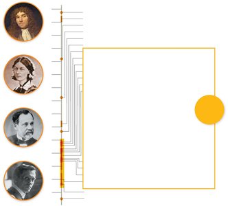
The development of microbiology as a science involved contributions from many scientists. The timeline includes the invention of microscopes, the establishment of aseptic techniques, and the formulation of the germ theory of disease.
Robert Hooke: First to publish descriptions of cells.
Antonie van Leeuwenhoek: Refined microscopes and first observed bacteria.
Louis Pasteur: Disproved spontaneous generation, developed pasteurization, and vaccines.
Robert Koch: Developed techniques for isolating bacteria and formulated Koch’s postulates.
Semmelweis, Lister, Nightingale: Pioneered aseptic techniques in healthcare.
What Is Microbiology?
Definition and Scope
Microbiology encompasses the study of cellular and noncellular microorganisms. It spans healthcare, agriculture, industry, and environmental sciences. Microbes are essential for food production, medication synthesis, and environmental remediation.
Cellular Microorganisms: Bacteria, archaea, protists, fungi, helminths.
Noncellular Entities: Viruses and prions.
Microbial Diversity: Microbes inhabit nearly every region of Earth.
Comparison of Living and Nonliving Agents Studied in Microbiology
Microbe | Cell Type | Notes |
|---|---|---|
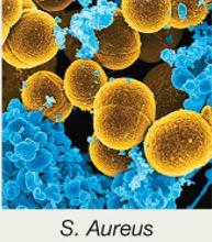
Bacteria | Prokaryotic | Unicellular; pathogenic and nonpathogenic |
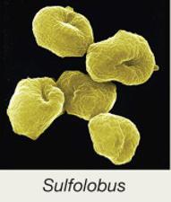
Archaea | Prokaryotic | Unicellular; nonpathogenic; extremophiles |
Protists | Eukaryotic | Unicellular/multicellular; pathogenic and nonpathogenic |
Fungi | Eukaryotic | Unicellular/multicellular; pathogenic and nonpathogenic |
Helminths | Eukaryotic | Multicellular; parasitic worms |
Viruses | Nonliving | Infect animal, plant, or bacterial cells; DNA or RNA genome |
Prions | Nonliving | Infectious proteins; transmitted by transplant or ingestion |
Spontaneous Generation Versus Biogenesis
Historical Debate and Experiments
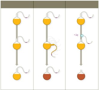
Scientists debated whether life originated from nonliving matter (spontaneous generation) or from existing life (biogenesis). Pasteur’s experiments with S-necked flasks provided evidence for biogenesis.
Spontaneous Generation: Life arises from nonliving items.
Biogenesis: Life emerges from existing life.
Pasteur’s Experiment: Demonstrated that microbes in air contaminate broth only when allowed to enter.
Germ Theory of Disease and Koch’s Postulates
Establishing Microbes as Disease Agents
The germ theory of disease states that microbes cause infectious diseases. Koch’s postulates provide a systematic method for identifying the causative agent of a disease.
Koch’s Postulates:
The same organism must be present in every case of the disease.
The organism must be isolated and grown as a pure culture.
The isolated organism should cause the same disease when inoculated into a susceptible host.
The organism must be re-isolated from the inoculated, diseased animal.
Hand Hygiene and Aseptic Techniques
Preventing Healthcare-Associated Infections
Aseptic techniques are essential in medical settings to prevent healthcare-acquired infections (HAIs). Key practices include hand washing, sterilizing instruments, and decontaminating surfaces.
Ignaz Semmelweis: Introduced hand washing to reduce childbed fever.
Joseph Lister: Developed aseptic surgery techniques.
Florence Nightingale: Established aseptic practices in nursing.
The Scientific Method in Microbiology
Principles and Application
The scientific method is the guiding principle for investigating questions in microbiology. It involves making observations, forming hypotheses, collecting and analyzing data, and drawing conclusions.
Observation: Data collected using senses or instruments.
Hypothesis: Proposed explanation for an observation.
Conclusion: Interpretation of data to support or refute the hypothesis.
Scientific Law vs. Theory: Laws predict what happens; theories explain how and why.
Classifying Microbes and Their Interactions
Taxonomic Hierarchy and Nomenclature
Microbes are classified using a hierarchical system from domain to species. Binomial nomenclature provides a standardized naming system.
Taxonomic Hierarchy: Domain, Kingdom, Phylum, Class, Order, Family, Genus, Species.
Three Domains: Bacteria, Archaea, Eukarya.
Six Kingdoms: Archaea, Bacteria, Fungi, Plantae, Animalia, Protists.
Binomial Nomenclature: Genus (capitalized) + species (lowercase), italicized (e.g., Escherichia coli).
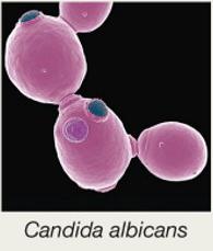
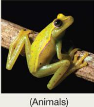
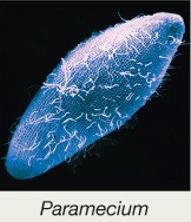
Kingdom | Domain | Example |
|---|---|---|
Archaea | Archaea |
|
Bacteria | Bacteria |
|
Fungi | Eukarya |
|
Plantae | Eukarya |
|
Animalia | Eukarya |
|
Protists | Eukarya |
|

Microbes and Symbiotic Relationships
Types of Interactions
Microbes interact with their hosts in various ways, including parasitism (harmful), mutualism (beneficial), and commensalism (neutral).
Parasitism: Microbe harms the host.
Mutualism: Both host and microbe benefit.
Commensalism: Microbe benefits without affecting the host.
Normal Microbiota and the Human Microbiome
Establishment and Functions
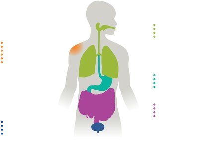
The human microbiome consists of bacteria, archaea, and eukaryotic microbes living in and on the body. These microbes train the immune system, produce vitamins, aid digestion, and may influence mood and brain function.
Colonization: Begins at birth and is influenced by delivery method and feeding.
Functions: Immune training, vitamin production, digestion, protection against pathogens.
Disruption: Antibiotic therapy can disturb normal microbiota, leading to opportunistic infections.
Biofilms
Formation and Healthcare Implications
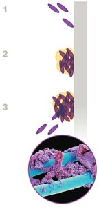
Biofilms are sticky communities of microbes that attach to surfaces and are highly resistant to antibiotics and immune responses. They are implicated in 60–80% of infectious diseases.
Formation: Microbes attach, secrete a matrix, and develop protective layers.
Healthcare Impact: Biofilms form on medical devices and are difficult to eradicate.
Growing, Staining, and Viewing Microbes
Culture Media and Aseptic Techniques
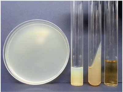
Microbes are grown in laboratory media such as broths, plates, slants, and deeps. Aseptic techniques are used to prevent contamination.
Culture Media: Mixtures of nutrients; agar is used as a solidifying agent.
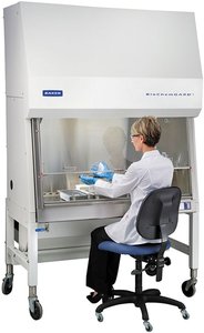
Aseptic Techniques: Sterile media, instruments, and protective clothing.
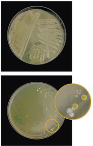
Streak Plate Technique: Used to isolate colonies for study.
Staining Techniques
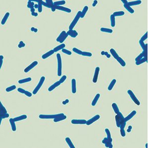
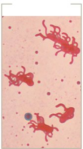
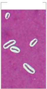
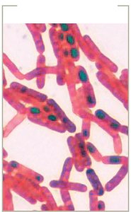
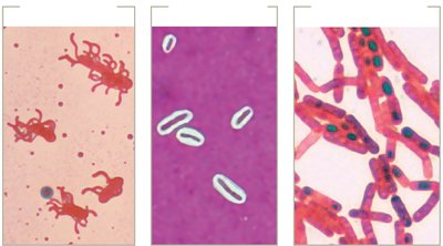
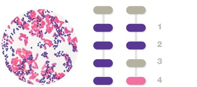
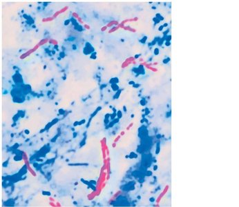
Stains increase contrast for microscopic viewing. Simple stains use one dye, while structural stains highlight features like flagella, capsules, and endospores. Differential stains, such as Gram and acid-fast, distinguish cell wall properties.
Simple Stains: Determine size, shape, and arrangement.
Structural Stains: Reveal flagella, capsules, and endospores.
Differential Stains: Gram stain (purple for Gram-positive, pink for Gram-negative), acid-fast stain (red for acid-fast, blue for non–acid-fast).
Microscopy in Microbiology
Types and Principles
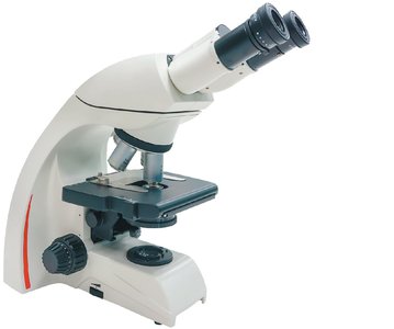
Microscopy is central to microbiology, allowing visualization of microbes. Light microscopy uses visible light, while electron microscopy uses electron beams for higher resolution.
Compound Light Microscope: Most common; uses ocular and objective lenses.
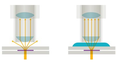
Resolution: Ability to distinguish two points as separate; improved with immersion oil.
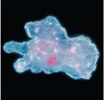
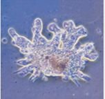
Types of Light Microscopy: Bright field, dark field, phase contrast, differential interference contrast.
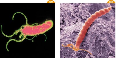
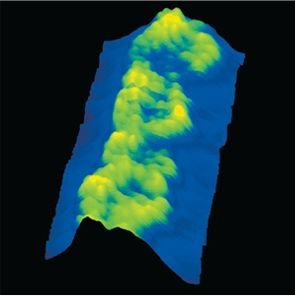
Electron Microscopy: Transmission (TEM) and scanning (SEM) provide high magnification and resolution.
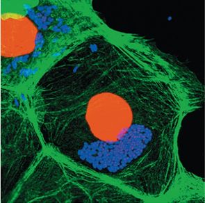
Fluorescence Microscopy: Uses fluorescent dyes for sensitive detection.
Summary and Key Takeaways
Microbiology studies a diverse range of organisms, both living and nonliving.
Historical experiments established biogenesis and the germ theory of disease.
Classification systems and nomenclature are essential for organizing microbial diversity.
Symbiotic relationships and the human microbiome are central to health and disease.
Biofilms pose significant challenges in healthcare.
Laboratory techniques for culturing, staining, and viewing microbes are foundational skills.
Microscopy enables detailed study of microbial structure and function.
