 뒤로
뒤로Foundations and Diversity of Microbiology: Key Concepts and Structures
스터디 가이드 - 스마트 노트
 자료에 맞춘 맞춤형 노트, 핵심 정의, 예시, 맥락을 확장해 제공합니다.
Introduction to Microbiology
What is Science?
Science is a systematic approach to understanding the natural world through observation, experimentation, and reasoning. It relies on the scientific method to generate knowledge that is testable and reproducible.
Scientific Method: Involves making observations, forming hypotheses, conducting experiments, and developing theories or laws based on consistent results.
Ockham’s Razor: The principle that the simplest explanation consistent with the facts is preferred.
What is Microbiology?
Microbiology is the study of microscopic organisms, including bacteria, viruses, fungi, protozoa, and algae. It explores their structure, function, classification, and role in health, disease, and the environment.
Branches of Microbiology: Bacteriology, Virology, Mycology, Parasitology, Phycology, Immunology.
History of Microbiology
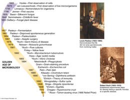
The field of microbiology has evolved through key discoveries and technological advancements, particularly during the "Golden Age of Microbiology." Early observations and experiments laid the foundation for understanding microorganisms and their impact on health and disease.
Milestones: Discovery of cells, development of vaccines, germ theory of disease, and identification of specific pathogens.
Disproving Spontaneous Generation
Key Experiments and Theories
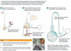
Spontaneous generation was the belief that life could arise from nonliving matter. This idea was disproven through carefully designed experiments, most notably by Louis Pasteur.
Pasteur’s Experiment: Used swan-neck flasks to show that microorganisms do not arise spontaneously but come from other microbes in the environment.
Controls: Included boiling broth, using straight and bent neck flasks, and observing microbial growth.
Golden Age of Microbiology
Major Discoveries and Contributors
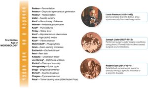
The Golden Age of Microbiology (mid-1800s to early 1900s) was marked by rapid advances in the identification of microbes and their roles in disease, fermentation, and immunity.
Louis Pasteur: Demonstrated fermentation, disproved spontaneous generation, developed vaccines, and introduced pasteurization.
Robert Koch: Established Koch’s postulates, linking specific microbes to specific diseases.
Joseph Lister: Introduced antiseptic techniques in surgery.
Koch’s Postulates
Establishing Microbial Etiology of Disease
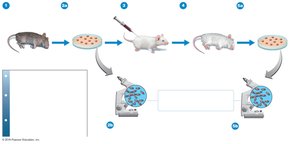
Koch’s postulates are a set of criteria used to prove that a specific microorganism causes a specific disease. They remain foundational in medical microbiology for identifying pathogens.
The same pathogen must be present in every case of the disease.
The pathogen must be isolated and grown in pure culture.
The cultured pathogen must cause disease when introduced into a healthy host.
The pathogen must be re-isolated from the experimentally infected host and shown to be the same as the original organism.
Exceptions: Some pathogens (e.g., Treponema pallidum for syphilis, HIV for AIDS) cannot be cultured or do not cause disease in all hosts.
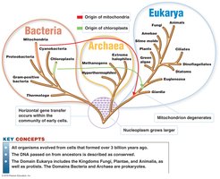
Three-Domain System and Evolutionary Relationships
Bacteria, Archaea, and Eukarya
All life forms are classified into three domains based on genetic and biochemical differences: Bacteria, Archaea, and Eukarya. This system reflects evolutionary relationships and the origins of key cellular structures.
Bacteria: Prokaryotic, peptidoglycan cell walls, ester-linked membrane lipids.
Archaea: Prokaryotic, no peptidoglycan, ether-linked membrane lipids, often extremophiles.
Eukarya: Eukaryotic, includes fungi, plants, animals, and protists.
Cell Structure and Diversity
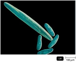
Size and Morphology of Microbial Cells
Microbial cells vary greatly in size and shape. Eukaryotic cells are generally larger (10–100 μm) than prokaryotic cells (0.2–2 μm), with viruses being much smaller (20–300 nm).
Volume Comparison: If a eukaryotic cell is 20 μm per side and a prokaryotic cell is 2 μm per side (assuming cubic shape), the eukaryote is times larger in volume.
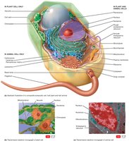
Eukaryotic Cell Structure
Eukaryotic cells contain membrane-bound organelles, including a nucleus, mitochondria, endoplasmic reticulum, and (in plants/algae) chloroplasts. These structures compartmentalize cellular functions.
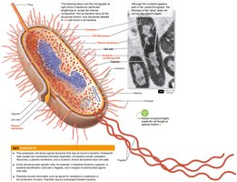
Prokaryotic Cell Structure
Prokaryotic cells lack a true nucleus and membrane-bound organelles. Key structures include the nucleoid, ribosomes, plasma membrane, cell wall, and sometimes capsules, flagella, and pili.
Shapes and Arrangements of Prokaryotic Cells
Cocci, Bacilli, and Other Forms
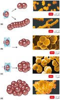
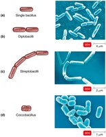
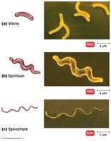
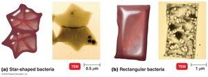
Bacteria exhibit a variety of shapes and arrangements, which are important for identification and classification.
Cocci: Spherical; may form pairs (diplococci), chains (streptococci), clusters (staphylococci), tetrads, or sarcinae.
Bacilli: Rod-shaped; may be single, paired (diplobacilli), chained (streptobacilli), or coccobacilli.
Spiral Forms: Vibrio (comma-shaped), spirillum (rigid spiral), spirochete (flexible spiral).
Unusual Shapes: Some bacteria are star-shaped or rectangular.
Cell Membrane & Transport
Structure and Function of the Plasma Membrane
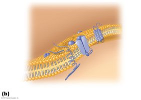
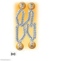
The plasma membrane is a selectively permeable barrier composed of a phospholipid bilayer with embedded proteins. It regulates the movement of substances into and out of the cell.
Phospholipid Bilayer: Hydrophilic heads face outward, hydrophobic tails face inward.
Proteins: Integral and peripheral proteins serve as channels, receptors, and enzymes.
Cell Wall Structure and Gram Staining
Gram-Positive vs. Gram-Negative Bacteria
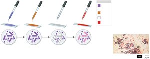
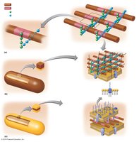
The cell wall provides shape and protection against osmotic lysis. Gram staining differentiates bacteria based on cell wall structure.
Gram-Positive: Thick peptidoglycan layer, retains crystal violet stain (purple).
Gram-Negative: Thin peptidoglycan layer, outer membrane with lipopolysaccharide (LPS), stains red with safranin.
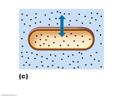
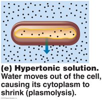
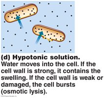
Osmosis and Cell Wall Function
Osmotic Environments
The cell wall helps maintain cell integrity in different osmotic environments.
Isotonic: No net water movement.
Hypertonic: Water leaves the cell, causing plasmolysis.
Hypotonic: Water enters the cell; strong walls prevent lysis, weak walls result in bursting.
Endospores
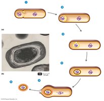
Formation and Resistance
Endospores are highly resistant, dormant structures formed by certain bacteria (e.g., Bacillus, Clostridium) to survive extreme conditions.
Resistance: Endospores withstand heat, chemicals, desiccation, radiation, and freezing.
Diseases: Endospore-formers cause anthrax, tetanus, botulism, and gas gangrene.
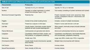
Principal Differences between Prokaryotic and Eukaryotic Cells
Comparison Table
The following table summarizes the main differences between prokaryotic and eukaryotic cells, including size, structure, and genetic organization.
Characteristic | Prokaryotic | Eukaryotic |
|---|---|---|
Size of Cell | Typically 0.2–2.0 μm | Typically 10–100 μm |
Nucleus | No true nucleus | True nucleus with nuclear membrane |
Membrane-Enclosed Organelles | Absent | Present (e.g., mitochondria, ER, Golgi) |
Cell Wall | Usually present; peptidoglycan | Present in plants/fungi (cellulose/chitin); absent in animals |
Ribosomes | 70S | 80S (cytoplasm), 70S (organelles) |
Chromosome (DNA) | Single, circular, haploid | Multiple, linear, diploid or more |
Cell Division | Binary fission | Mitotic division |
Eukaryotic Microorganisms
Fungi
Fungi include molds (multicellular, aerobic) and yeasts (unicellular, facultative anaerobes). They play important roles in decomposition, food production, and as sources of antibiotics.
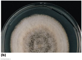
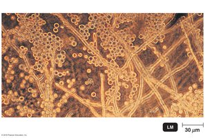
Molds: Form hyphae and spores; visible as colonies on agar.
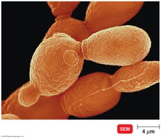
Yeasts: Reproduce by budding; important in fermentation.
Dimorphism: Some fungi can exist as both yeast and mold forms.
Protozoa
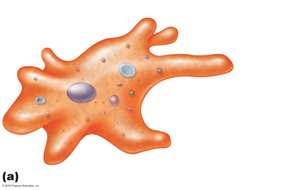
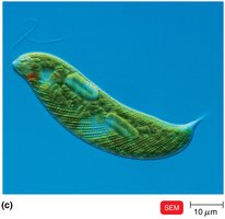
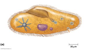
Protozoa are single-celled eukaryotes classified by their motility structures: pseudopodia (amoebae), flagella (euglenoids), or cilia (ciliates). They are important in food chains and as pathogens.
Amoebae: Move by pseudopodia.
Euglenoids: Move by flagella; some are photosynthetic.
Ciliates: Covered with cilia for movement and feeding.
Algae
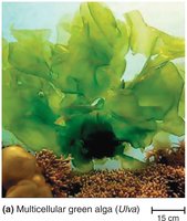
Algae are photosynthetic eukaryotes found in aquatic environments. They produce oxygen and serve as the base of many food webs. Some are used industrially (e.g., agar production).
Green Algae: Example: Ulva, multicellular and sheet-like.
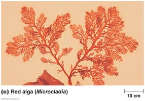
Red Algae: Example: Microcladia, with branching fronds.
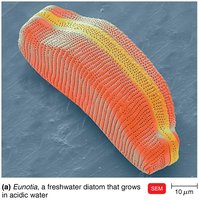
Diatoms: Unicellular algae with silica cell walls, important in aquatic ecosystems.
Summary
This guide covers foundational concepts in microbiology, including the scientific method, history and milestones, cell structure, classification, and the diversity of microbial life. Understanding these principles is essential for further study in microbial physiology, genetics, and pathogenesis.
