 뒤로
뒤로Functional Anatomy of Prokaryotic and Eukaryotic Cells: Structure and Function
스터디 가이드 - 스마트 노트
 자료에 맞춘 맞춤형 노트, 핵심 정의, 예시, 맥락을 확장해 제공합니다.
Functional Anatomy of Prokaryotic and Eukaryotic Cells
Introduction
This section explores the structural and functional differences between prokaryotic and eukaryotic cells, focusing on their cellular components, shapes, arrangements, and specialized structures. Understanding these differences is fundamental to microbiology, as it underpins the classification, physiology, and pathogenicity of microorganisms.
Prokaryotic and Eukaryotic Cells
Definitions and Key Differences
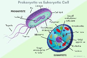
Prokaryote: Derived from Greek for "prenucleus"; organisms whose cells lack a true nucleus and membrane-bound organelles. Includes Bacteria and Archaea.
Eukaryote: Derived from Greek for "true nucleus"; organisms whose cells possess a nucleus enclosed by a membrane and various membrane-bound organelles. Includes Fungi, Algae, Protozoa, Plants, and Animals.
Bacterial Cell Shapes and Arrangements
Basic Shapes
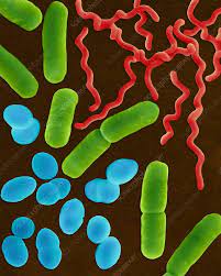
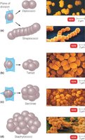
Coccus: Spherical-shaped bacteria.
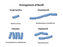
Bacillus: Rod-shaped bacteria. The term also refers to the genus Bacillus.
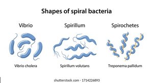
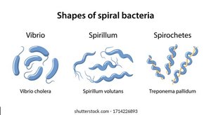
Spiral Forms: Includes vibrio (comma-shaped), spirillum (rigid spiral), and spirochete (flexible spiral).
Most bacteria are monomorphic (single shape), but some are pleomorphic (variable shapes).
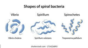
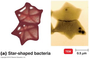
Unusual Bacterial Shapes
Star-shaped bacteria and rectangular bacteria are rare morphological variants.
Structure of a Prokaryotic Cell
General Anatomy
Key structures include the cell wall, plasma membrane, cytoplasm, nucleoid, ribosomes, inclusions, and external appendages (flagella, fimbriae, pili).
Glycocalyx
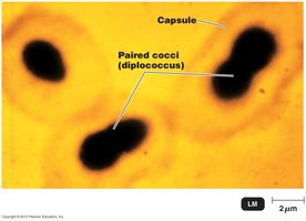
The glycocalyx is a viscous, gelatinous outer layer composed mainly of polysaccharides, sometimes with proteins or lipids.
Two main forms:
Capsule: Organized, dense, and firmly attached to the cell wall; often increases pathogenicity by protecting against phagocytosis.
Slime layer: Unorganized, loose, and easily removed.
Functions:
Protection: Shields against dehydration, antibiotics, chemicals, and immune defenses.
Adhesion: Facilitates attachment to surfaces and formation of biofilms.
Flagella
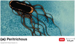
Flagella are long, whip-like appendages used for motility.
Structure: Composed of flagellin protein, attached to the cell by a basal body and hook.
Arrangements: Monotrichous (single), lophotrichous (tuft), amphitrichous (both ends), peritrichous (all over).
Movement: "Run and tumble" pattern; taxis (movement toward/away from stimuli).
Flagellar proteins serve as H antigens (e.g., E. coli O157:H7).
Fimbriae and Pili
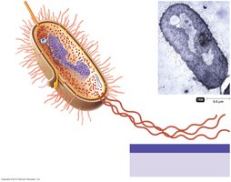
Fimbriae: Short, hairlike structures for attachment; important in biofilm formation and colonization (e.g., E. coli in intestines).
Pili: Longer than fimbriae; usually one or two per cell; involved in DNA transfer (conjugation or sex pilus).
Cell Wall
Provides shape, protection from osmotic lysis, and serves as an anchor for flagella.
Composed mainly of peptidoglycan in bacteria.
Peptidoglycan Structure
Polymer of alternating N-acetylglucosamine (NAG) and N-acetylmuramic acid (NAM) sugars, cross-linked by short peptides.
Gram-Positive vs. Gram-Negative Cell Walls
Feature | Gram-Positive | Gram-Negative |
|---|---|---|
Peptidoglycan | Thick, multilayered | Thin, single layer |
Teichoic acids | Present | Absent |
Outer membrane | Absent | Present (contains LPS) |
Lipid A (endotoxin) | Absent | Present |
Sensitivity to lysozyme/penicillin | High | Low |
Gram Stain Mechanism
Gram-positive: Alcohol dehydrates peptidoglycan, trapping crystal violet-iodine complex.
Gram-negative: Alcohol dissolves outer membrane, allowing dye to wash out.
Atypical Cell Walls
Acid-fast bacteria (e.g., Mycobacterium): Waxy mycolic acid layer in addition to peptidoglycan.
Plasma Membrane
Phospholipid bilayer with embedded proteins; selectively permeable.
Functions: Transport, energy generation, photosynthesis (in some bacteria), and cell signaling.
Transport mechanisms:
Simple diffusion: Movement from high to low concentration.
Facilitated diffusion: Uses transporter proteins.
Active transport: Requires energy (ATP or PEP).
Group translocation: Substance is chemically modified during transport (unique to prokaryotes).
Cytoplasm and Internal Structures
Cytoplasm: Gel-like matrix containing water, enzymes, nutrients, wastes, and cell structures.
Nucleoid: Region containing the bacterial chromosome (circular, double-stranded DNA).
Plasmids: Small, circular DNA molecules; carry non-essential but advantageous genes (e.g., antibiotic resistance).
Ribosomes: Sites of protein synthesis; prokaryotic ribosomes are 70S (50S + 30S subunits).
Inclusions: Reserve deposits (e.g., magnetosomes, gas vacuoles).
Endospores: Dormant, highly resistant structures formed by some Gram-positive bacteria (e.g., Bacillus, Clostridium).
Eukaryotic Cell Structure
Flagella and Cilia
Flagella: Few, long projections for movement.
Cilia: Numerous, short projections for movement or moving substances along surfaces.
Both have a "9+2" arrangement of microtubules and are anchored by a basal body.
Cell Wall and Glycocalyx
Most eukaryotes have a cell wall (composition varies: cellulose in plants, chitin in fungi, glucan/mannan in yeast).
Animal cells lack a cell wall but have a glycocalyx for protection and cell recognition.
No peptidoglycan in eukaryotic cell walls; thus, antibiotics targeting peptidoglycan are not harmful to host cells.
Plasma Membrane and Transport
Similar structure to prokaryotes but may contain sterols for rigidity.
Transport mechanisms include simple diffusion, facilitated diffusion, active transport, and endocytosis (phagocytosis and pinocytosis).
Cytoplasm and Organelles
Cytoplasm: Area between plasma membrane and nucleus; contains cytosol, cytoskeleton, and organelles.
Nucleus: Contains DNA packaged as chromatin; surrounded by a nuclear envelope with pores.
Ribosomes: 80S (60S + 40S subunits) in cytoplasm and on rough ER; 70S in mitochondria and chloroplasts.
Endoplasmic Reticulum (ER): Rough ER (with ribosomes) synthesizes proteins; smooth ER synthesizes lipids.
Golgi Complex: Modifies, sorts, and packages proteins and lipids for secretion or delivery to other organelles.
Lysosomes: Contain digestive enzymes for breaking down macromolecules and pathogens.
Vacuoles: Storage and support (especially in plants).
Mitochondria: Site of cellular respiration and ATP production.
Chloroplasts: Site of photosynthesis in plants and algae.
Peroxisomes: Break down fatty acids and detoxify harmful substances.
Centrosomes: Organize microtubules and are important in cell division.
Endosymbiotic Theory
Proposes that eukaryotic organelles (mitochondria, chloroplasts) originated from prokaryotic cells engulfed by ancestral eukaryotes.
Supported by similarities in DNA, ribosomes, and reproduction between these organelles and prokaryotes.
