 뒤로
뒤로Introduction to Microbiology: Core Concepts and Historical Foundations
스터디 가이드 - 스마트 노트
 자료에 맞춘 맞춤형 노트, 핵심 정의, 예시, 맥락을 확장해 제공합니다.
Introduction to Microbiology
What Is Microbiology?
Microbiology is the scientific study of microorganisms, or microbes, which are typically too small to be seen with the naked eye. This field encompasses a diverse range of organisms and entities, both living and nonliving.
Microbes include cellular, living microorganisms such as bacteria, archaea, fungi, protists, and helminths.
Nonliving/noncellular entities studied in microbiology include viruses and prions (infectious proteins).
Some microbes, such as certain fungi, helminths, and protists, are not always microscopic but have microscopic life stages.
Major Groups of Microbes
Microorganisms are classified into several major groups based on their cellular structure and life cycle.
Bacteria: Unicellular prokaryotes found in diverse environments.
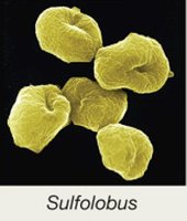
Archaea: Unicellular prokaryotes, often found in extreme environments; no known pathogens.
Fungi: Includes unicellular (yeasts) and multicellular (molds, mushrooms) organisms.
Protists: Diverse group including protozoa and algae.
Helminths: Parasitic worms, some of which have microscopic stages.
Viruses: Nonliving infectious agents composed of genetic material and protein.
Prions: Infectious proteins lacking nucleic acids.
Table: Living and Nonliving Agents Studied in Microbiology
Type | Cellular Structure | Examples |
|---|---|---|
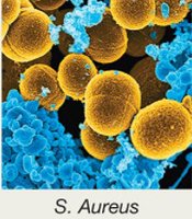
Bacteria | Unicellular, prokaryotic | Escherichia coli, Staphylococcus aureus |
Archaea | Unicellular, prokaryotic | Sulfolobus |
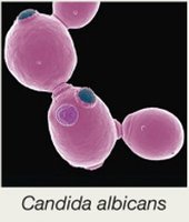
Fungi | Unicellular or multicellular, eukaryotic | Candida albicans |
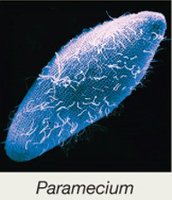
Protists | Unicellular or multicellular, eukaryotic | Paramecium |
Helminths | Multicellular, eukaryotic | Tapeworms, roundworms |
Viruses | Noncellular | Influenza virus, HIV |
Prions | Noncellular (protein only) | Prion causing Creutzfeldt-Jakob disease |
Cell Types and Evolution
Prokaryotic vs. Eukaryotic Cells
Microorganisms can be classified based on their cellular organization:
Prokaryotic cells: Lack a nucleus and membrane-bound organelles; include bacteria and archaea. Evolved about 3.5 billion years ago and are considered the earliest life forms.
Eukaryotic cells: Possess a nucleus and membrane-bound organelles; include all multicellular organisms and many unicellular organisms (e.g., amoebae, yeast).
The endosymbiotic theory explains the origin of eukaryotic organelles such as mitochondria and chloroplasts from ancestral prokaryotes.
Microbiology in Society
Applications and Importance
Microbiology spans a wide variety of fields, including healthcare, agriculture, industry, and environmental sciences. Humans rely on microbes for:
Food production (e.g., fermentation)
Making medications (e.g., antibiotics, vaccines)
Breaking down environmental hazards (bioremediation)
Microbes and Disease
Pathogens and Human Health
Pathogens are microbes that cause disease. Of the millions of microbial species, only about 1,400 are known to infect humans, representing less than 1% of all microbes.
True pathogens: Always cause disease in humans.
Opportunistic pathogens: Cause disease only in weakened hosts.
Historical Foundations of Microbiology
The Golden Age of Microbiology (1850–1920)
This era saw major advances in microscopy, microbial isolation, and cultivation techniques, laying the foundation for modern microbiology.
Spontaneous Generation vs. Biogenesis
Spontaneous generation: The hypothesis that life arises from nonliving matter.
Biogenesis: The principle that life arises only from pre-existing life.
Key contributors:
Robert Hooke: First to publish descriptions of cells.
Antonie van Leeuwenhoek: Improved microscopes and first observed bacteria.
Louis Pasteur: Demonstrated biogenesis, developed pasteurization, and created vaccines for anthrax and rabies.
Germ Theory of Disease
The germ theory states that microbes cause infectious diseases. Robert Koch developed methods to identify the specific causative agents of diseases, including:
Staining techniques
Media for bacterial isolation and cultivation
Koch's postulates for linking microbes to diseases
Koch’s Postulates
The same organism must be present in every case of the disease.
The organism must be isolated from the diseased host and grown as a pure culture.
The isolated organism should cause the same disease when inoculated into a susceptible host.
The organism must be re-isolated from the inoculated, diseased animal.
Aseptic Techniques and Infection Control
Development of Aseptic Techniques
Several pioneers emphasized the importance of aseptic techniques in medical settings to prevent infection:
Ignaz Semmelweis: Advocated hand washing to reduce childbed fever.
Joseph Lister: Introduced sterilization of instruments and antiseptic surgery.
Florence Nightingale: Established aseptic techniques in nursing.
Aseptic processes prevent healthcare-acquired infections (HAIs) and include hand washing, wearing gloves, sterilizing instruments, and decontaminating surfaces.
Taxonomy and Classification
Taxonomic Hierarchy
Taxonomy is the science of classifying organisms based on shared features. The taxonomic hierarchy includes eight ranks:
Domain
Kingdom
Phylum
Class
Order
Family
Genus
Species
Mnemonic: "Delightful King Philip came over for great spaghetti"
Three-Domain and Six-Kingdom Systems
Three domains: Bacteria, Archaea, Eukarya
Six kingdoms: Bacteria, Archaea, Protista, Fungi, Plantae, Animalia
Domain | Kingdom | Example |
|---|---|---|
Bacteria | Bacteria | S. aureus |
Archaea | Archaea | Sulfolobus |
Eukarya | Protista | Paramecium |
Eukarya | Fungi | Candida albicans |
Eukarya | Plantae | Plants |
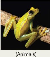
Eukarya | Animalia | Animals |

Scientific Names
Carl Linnaeus established the binomial nomenclature system, which uses two names: the genus (capitalized) and the species (lowercase), both italicized (e.g., Escherichia coli).
Host–Microbe Interactions
Symbiotic Relationships
Microbes and humans engage in various symbiotic relationships:
Parasitism: Microbe harms the host.
Mutualism: Both host and microbe benefit.
Commensalism: Microbe benefits without affecting the host.
Pathogens are considered to have a parasitic relationship with their host.
Normal Microbiota and the Human Microbiome
Human Microbiome Project
The Human Microbiome Project aims to characterize all microbes living in and on the human body. Major sites include the skin, nose, mouth, gut, and genital/urinary tract.
Functions of Normal Microbiota
Train the immune system
Produce vitamins
Aid in digestion
May influence mood and brain function
Establishment and Disruption of Normal Microbiota
Babies are colonized during delivery and early interactions.
Factors such as delivery method and feeding influence microbiota development.
Antibiotic therapy can disrupt normal microbiota, increasing risk for opportunistic infections (e.g., Candida albicans overgrowth).
Transient Microbiota
Transient microbiota are temporary and do not persist as stable residents. They can be removed by proper hygiene.
Biofilms
Formation and Importance
Biofilms are structured communities of microbes attached to surfaces and embedded in a self-produced matrix. They can form on teeth, medical devices, and other surfaces.
Biofilms are more resistant to antibiotics and immune responses.
Estimated to cause 60–80% of human infectious diseases.
Microbial Cultivation and Identification
Growth Media
Growth media are nutrient mixtures used to culture microbes in the laboratory. Types include broths, plates, slants, and deeps. Agar is often used as a solidifying agent.
Aseptic Culture Techniques
Aseptic techniques are essential for obtaining pure cultures and preventing contamination. Methods include using sterile media, instruments, and protective clothing.
Biological safety cabinets protect both the culture and the researcher.
Streak plate technique is used to isolate colonies.
Gram Stain
Principle and Procedure
The Gram stain differentiates bacteria into Gram-positive (purple) and Gram-negative (pink) based on cell wall structure.
Crystal violet (primary stain)
Iodine (mordant)
Acetone-alcohol (decolorizer)
Safranin (counterstain)
Gram-positive bacteria have thick peptidoglycan layers; Gram-negative bacteria have thin peptidoglycan and an outer membrane rich in lipids.
Interpretation and Errors
Over-decolorization can cause Gram-positive cells to appear Gram-negative.
Fresh cultures (24–48 hours old) minimize errors.
Microscopy in Microbiology
Light Microscopy
Light microscopes use visible light and lenses to magnify specimens. The compound light microscope is the most common type.
Electron Microscopy
Electron microscopes use electron beams for much higher resolution imaging than light microscopes.
Resolution improves with smaller wavelengths (electron beams: 1 nm; visible light: 400 nm).
Electron microscopy is expensive and requires specialized training.
Additional info: This guide covers foundational concepts from Chapter 1 of a college-level microbiology course, including definitions, historical context, and essential laboratory techniques.
