뒤로
뒤로Introduction to Microbiology: Foundations, Methods, and Applications
스터디 가이드 - 스마트 노트
 자료에 맞춘 맞춤형 노트, 핵심 정의, 예시, 맥락을 확장해 제공합니다.
Introduction to Microbiology
Definition and Scope
Microbiology is the study of microorganisms (microbes), which are organisms too small to be seen with the naked eye. This field encompasses a wide range of life forms, including bacteria, archaea, fungi, protists, helminths, viruses, and prions. Microbiology is foundational to healthcare, agriculture, industry, and environmental sciences.
Microbe: Any microscopic organism, including both cellular (e.g., bacteria, fungi) and noncellular entities (e.g., viruses, prions).
Applications: Food production, medication synthesis, environmental remediation, and more.
Types of Microorganisms
Cellular, living microorganisms: Bacteria, archaea, fungi, protists, helminths.
Nonliving/noncellular entities: Viruses, prions.
Some microbes, such as certain fungi and helminths, are not always microscopic but have microscopic life stages.
Prokaryotic vs. Eukaryotic Cells
Prokaryotic cells: Unicellular organisms lacking a nucleus (e.g., bacteria, archaea).
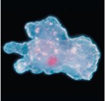
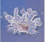
Eukaryotic cells: Organisms with a nucleus, including multicellular and some unicellular forms (e.g., amoebae, yeast).
Endosymbiotic theory: Explains the origin of eukaryotic organelles from prokaryotic ancestors.
A Brief History of Microbiology
Key Historical Figures and Events
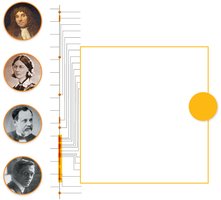
Robert Hooke: First to publish descriptions of cells.
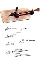
Antonie van Leeuwenhoek: Improved the microscope and first observed bacteria.
Carl Linnaeus: Developed the taxonomic naming system.
Louis Pasteur: Disproved spontaneous generation, developed pasteurization, and created vaccines for anthrax and rabies.
Robert Koch: Established the germ theory of disease and Koch's postulates.
Ignaz Semmelweis, Joseph Lister, Florence Nightingale: Pioneered aseptic techniques in healthcare.
Spontaneous Generation vs. Biogenesis
Debate over the origin of life led to two main theories:
Spontaneous generation: Life arises from nonliving matter.
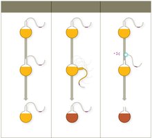
Biogenesis: Life arises from pre-existing life. Pasteur's S-necked flask experiment provided strong evidence for biogenesis.
Microbes and Disease
Pathogens and Opportunistic Pathogens
Pathogen: A microbe that causes disease. Less than 1% of microbes are pathogenic.
Opportunistic pathogen: Causes disease only in weakened hosts.
Germ Theory of Disease and Koch's Postulates
Germ theory: Microbes are the cause of infectious diseases.
Koch's postulates: Criteria to establish a causative relationship between a microbe and a disease:
The same organism must be present in every case of the disease.
The organism must be isolated and grown as a pure culture.
The isolated organism should cause the same disease in a healthy host.
The organism must be re-isolated from the experimentally infected host.
Aseptic Techniques and Hand Hygiene
Development of Aseptic Techniques
Ignaz Semmelweis: Advocated hand washing to prevent childbed fever.
Joseph Lister: Introduced sterilization of instruments and antiseptics in surgery.
Florence Nightingale: Established aseptic practices in nursing.
Aseptic techniques include hand washing, wearing gloves, sterilizing instruments, and decontaminating surfaces to prevent healthcare-associated infections (HAIs).
The Scientific Method in Microbiology
Steps of the Scientific Method
Formulate a question.
Propose a hypothesis.
Collect and analyze data (observations).
Draw a conclusion based on the data.
Observation: Data collected using senses or instruments. Conclusion: Interpretation of observations.
Scientific law: Predicts what happens (often mathematical). Scientific theory: Explains how and why something happens, supported by extensive evidence.
Taxonomy and Classification of Microbes
Taxonomic Hierarchy
Taxonomy is the science of classifying organisms based on shared features. The hierarchy from broadest to most specific is:
Domain
Kingdom
Phylum
Class
Order
Family
Genus
Species
Mnemonic: "Delightful King Philip came over for great spaghetti"
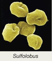
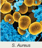
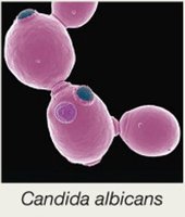
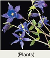
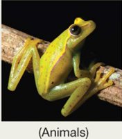
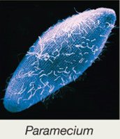
Three domains: Bacteria, Archaea, Eukarya.
Domain | Example |
|---|---|
Bacteria | Unicellular prokaryotes |
Archaea | Often extremophiles, no known pathogens |
Eukarya | Unicellular and multicellular eukaryotes |
Scientific Names
Binomial nomenclature: Two-part scientific name (Genus species), e.g., Escherichia coli.
Genus is capitalized, species is lowercase, both italicized.
Host–Microbe Interactions
Types of Symbiotic Relationships
Parasitism: Microbe harms the host.
Mutualism: Both host and microbe benefit.
Commensalism: Microbe benefits, host is unaffected.
Normal Microbiota and the Human Microbiome
The normal microbiota (normal flora) consists of bacteria, archaea, and eukaryotic microbes that inhabit the human body. They play roles in immune training, vitamin production, digestion, and possibly mood regulation.
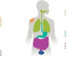
Normal microbiota can include potential pathogens, but usually do not cause disease due to competition and immune regulation.
Disruption (e.g., by antibiotics) can lead to opportunistic infections.
Establishment and Disruption of Microbiota
Babies acquire microbiota during birth and early life (influenced by delivery mode and feeding).
Antibiotic therapy can disrupt normal microbiota, allowing opportunistic pathogens to cause disease (e.g., Candida albicans yeast infections, antibiotic-associated diarrhea).
Biofilms
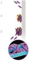
Biofilms are structured communities of microbes attached to surfaces and encased in a self-produced matrix. They are highly resistant to antibiotics and immune responses.
Common on teeth (dental plaque), medical devices, and water systems.
Microbial Growth and Laboratory Techniques
Growth Media
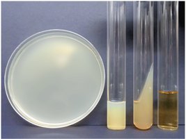
Growth media are nutrient mixtures used to culture microbes in the lab. Types include broths, agar plates, slants, and deeps.
Aseptic Culture Techniques
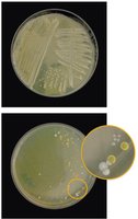
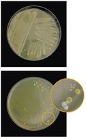
Pure cultures are isolated using aseptic techniques to prevent contamination.
Streak plate method is used to isolate colonies.
Microscopy and Staining
Staining Techniques
Simple stains: Use one dye to reveal cell size, shape, and arrangement.
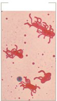
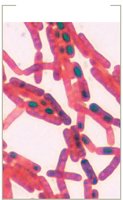
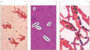
Structural stains: Highlight specific structures (flagella, capsules, endospores).
Flagella stain: Uses mordants to visualize flagella.
Capsule stain: Uses both acidic and basic dyes; capsules appear as halos.
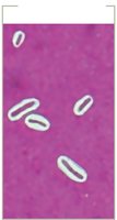
Endospore stain: Malachite green stains spores, safranin stains cells.
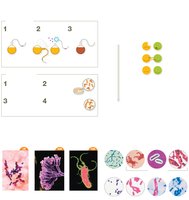
Summary of structural stains:
Differential stains: Distinguish between cell types.
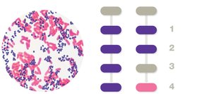
Gram stain: Differentiates Gram-positive (purple) and Gram-negative (pink) bacteria.
Acid-fast stain: Identifies bacteria with waxy cell walls (e.g., Mycobacterium).
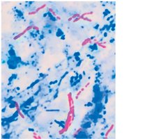
Microscopy
Light microscopy: Uses visible light and lenses to magnify specimens. The compound light microscope is most common.
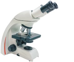
Resolution: Ability to distinguish two points as separate; typical light microscope resolution is about 200 nm.
Oil immersion: Used to improve resolution at high magnification by reducing light scattering.
Types of light microscopy: Bright field, dark field, phase contrast, differential interference contrast.
Electron and Advanced Microscopy
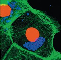
Electron microscopy: Uses electron beams for high-resolution imaging.
Transmission electron microscope (TEM): Views internal structures.
Scanning electron microscope (SEM): Views surface details.
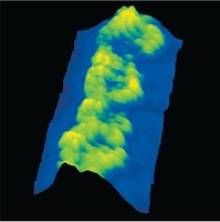
Other techniques: UV light microscopy, probe microscopy.
Visual Summary