 뒤로
뒤로Chapter 1
스터디 가이드 - 스마트 노트
 자료에 맞춘 맞춤형 노트, 핵심 정의, 예시, 맥락을 확장해 제공합니다.
Introduction to Microbiology
Definition and Scope
Microbiology is the study of microorganisms, or microbes, which are often invisible to the naked eye. The field encompasses both cellular (bacteria, archaea, fungi, protists, helminths) and acellular/noncellular entities (viruses, viroids, prions). Microbiology spans healthcare, agriculture, industry, and environmental sciences, highlighting the essential roles microbes play in food production, medication synthesis, and environmental remediation.
Historical Development
The timeline of microbiology includes key discoveries and figures such as Robert Hooke, Antonie van Leeuwenhoek, Louis Pasteur, Robert Koch, and Florence Nightingale. These pioneers contributed to the understanding of cell theory, biogenesis, germ theory of disease, and aseptic techniques. 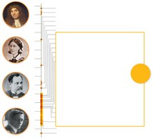
Microbes and Disease
Pathogens are microbes that cause disease, but less than 1% of all microbes are pathogenic. Pathogens can be classified as true pathogens (always cause disease) or opportunistic pathogens (cause disease in weakened hosts).
Disproving Spontaneous Generation
Biogenesis vs. Spontaneous Generation
Early debates centered on whether life arose from nonliving matter (spontaneous generation) or from existing life (biogenesis). Louis Pasteur's experiments with S-necked flasks demonstrated that airborne microbes, not spontaneous generation, were responsible for contamination. 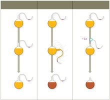
Germ Theory of Disease
Koch’s Postulates
Robert Koch established a systematic method to link specific microbes to diseases, known as Koch’s postulates:
The same organism must be present in every case of the disease.
The organism must be isolated and grown as a pure culture.
The isolated organism should cause the same disease when inoculated into a susceptible host.
The organism must be re-isolated from the inoculated, diseased animal.
Aseptic Techniques and Hand Hygiene
Contributions to Healthcare
Ignaz Semmelweis, Joseph Lister, and Florence Nightingale emphasized the importance of aseptic techniques, including hand washing, sterilizing instruments, and decontaminating surfaces, to prevent healthcare-acquired infections.
The Scientific Method in Microbiology
Principles and Application
The scientific method involves making observations, proposing hypotheses, collecting and analyzing data, and drawing conclusions. It is fundamental to investigating disease origins and treatments.
Law vs. Theory
Scientific Law: A precise statement or mathematical formula predicting a specific occurrence.
Scientific Theory: A hypothesis proven through repeated studies with consistent supporting conclusions.
Classifying Microbes and Their Interactions
Taxonomic Hierarchy
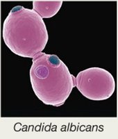
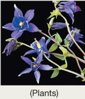
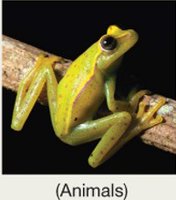
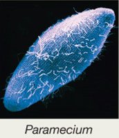
Organisms are classified from domain to species. The three domains are Bacteria, Archaea, and Eukarya. The six-kingdom system includes Animalia, Plantae, Fungi, Protista, Archaea, and Bacteria. 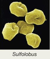
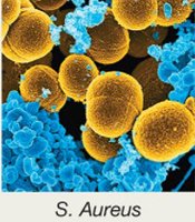
Binomial Nomenclature
Scientific names use a two-name system: Genus (capitalized) and species (lowercase), italicized (e.g., Escherichia coli).
Symbiotic Relationships
Parasitism: Hurts the host
Mutualism: Helps the host
Commensalism: No perceived benefit or cost to the host
Normal Microbiota and the Human Microbiome
Establishment and Functions
The human microbiome consists of bacteria, archaea, and eukaryotic microbes living in and on the body. Functions include training the immune system, producing vitamins, aiding digestion, and influencing mood and brain function. 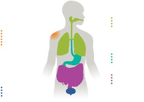
Disruptions and Transient Microbiota
Antibiotic therapy can disrupt normal microbiota, increasing infection risk. Transient microbiota are temporary and can be removed through hygiene.
Biofilms
Formation and Healthcare Implications
Biofilms are sticky communities of microbes attached to surfaces, protected by a matrix. They are resistant to antibiotics and immune responses, and are implicated in 60–80% of infectious diseases. 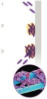
Growing, Staining, and Viewing Microbes
Culture Media
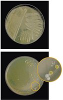
Growth media are mixtures of nutrients supporting microbial growth. Types include broths, plates, slants, and deeps. 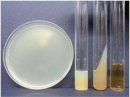
Aseptic Culture Techniques
Aseptic techniques prevent contamination in laboratory settings. The streak plate technique isolates colonies for study. 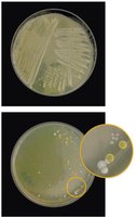
Staining Techniques
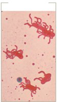
Simple stains: Use one dye to determine size, shape, and arrangement.
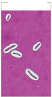
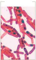
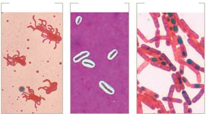
Structural stains: Reveal specific structures like flagella, capsules, and endospores.
Differential stains: Distinguish between cell types (e.g., Gram and acid-fast stains).
Gram Stain
The Gram stain differentiates bacteria into Gram-positive (purple) and Gram-negative (pink) based on cell wall structure. 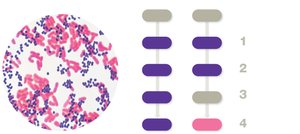
Acid-Fast Stain
Acid-fast staining distinguishes cells with waxy cell walls (e.g., Mycobacterium species). 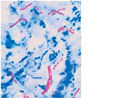
Microscopy
Compound Light Microscope
Light microscopy uses visible light and lenses to magnify specimens. The compound light microscope is the most common. 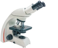
Oil Immersion
Immersion oil increases resolution by matching the refractive index of glass, preventing light scattering. 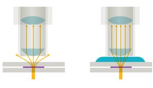
Types of Light Microscopy
Bright Field
Dark Field
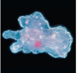
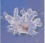
Phase Contrast
Differential Interference Contrast
Electron Microscopy
Electron microscopes use electron beams for high-resolution imaging.
Transmission Electron Microscopy (TEM): Provides 2D images of internal structures.
Scanning Electron Microscopy (SEM): Provides 3D images of surfaces.
Fluorescence Microscopy
Fluorescent dyes (fluorochromes) are used to stain samples, which emit visible light when illuminated by UV light. Immunofluorescence links dyes to antibodies for specific identification. 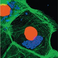
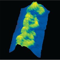
Visual Summary
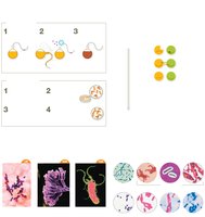
Additional info:
Microbiology is foundational for understanding infectious diseases, environmental processes, and biotechnology.
Laboratory techniques such as staining and microscopy are essential for identifying and classifying microbes.
Symbiotic relationships and the human microbiome are critical for health and disease prevention.
