뒤로
뒤로Chapter 3
스터디 가이드 - 스마트 노트
 자료에 맞춘 맞춤형 노트, 핵심 정의, 예시, 맥락을 확장해 제공합니다.
Prokaryotic Cell Basics
Overview of Prokaryotes
Prokaryotic cells are unicellular organisms that lack a membrane-bound nucleus and organelles. They are classified into two domains: Bacteria and Archaea. Life on Earth likely originated as prokaryotic cells about 3.8 billion years ago.
Bacteria and Archaea are distinct domains, each with unique cellular features.
Prokaryotes are fundamentally different from Eukaryotes, which possess a nucleus and organelles.
Prokaryotic cells lack a membrane-bound nucleus and organelles.
Classification and Evolution
Prokaryotes are divided into two domains: Bacteria and Archaea.
Eukaryotes form a separate domain.
All three domains share a common ancestor.

Prokaryotic Cell Structure
General Structure
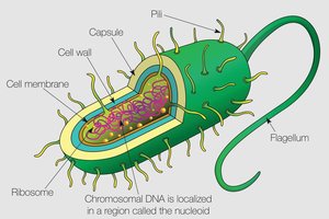
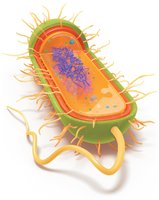
Prokaryotic cells have a simple structure but possess specialized features for survival and adaptation.
Plasma membrane: Selectively permeable lipid bilayer.
Cell wall: Provides rigidity and protection.
Capsule: Some cells have an additional protective layer.
Ribosomes: Sites of protein synthesis.
Nucleoid region: Location of chromosomal DNA.
Fimbriae and Flagella: Structures for adhesion and motility.
Sizes, Shapes, and Arrangements
Prokaryotes exhibit a wide range of sizes, shapes, and cellular arrangements, which are important for identification and pathogenicity.
Sizes range from very small (e.g., Mycoplasma) to very large (e.g., Thiomargarita namibiensis).
Most nutrients are obtained by diffusion, limiting cell size due to surface area-to-volume ratio.

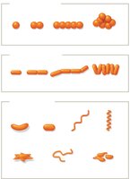
Shapes include: Bacillus (rod-shaped), Coccus (spherical), Vibrio (comma-shaped), Stella (star-shaped), Coccobacillus (ovoid), Spirochete (spiral-shaped).
Arrangements: Diplococci (paired cocci), Streptococci (chains), Staphylococci (clusters), Diplobacilli (paired rods), Streptobacilli (chains), Palisades (clusters of bacilli).
Cell Division: Binary Fission
Prokaryotic cells reproduce by binary fission, a simple and efficient process.
DNA is replicated.
Cell elongates.
Chromosomes are drawn to opposite ends.
Septum forms and partitions the cell.
Daughter cells separate.
Cell Envelope: Plasma Membrane and Cell Wall
Plasma Membrane Structure and Function
The plasma membrane is a thin, flexible, phospholipid bilayer that acts as a selective barrier.
Contains proteins that function as transporters, anchors, receptors, and enzymes.
Site for metabolic reactions, including ATP synthesis.
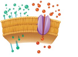
Exhibits selective permeability: small, noncharged molecules diffuse freely; ions and large polar molecules require transport proteins.
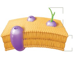
Membrane Fluidity
Membrane fluidity is essential for cell function and is influenced by temperature and lipid composition.
Warmer temperatures increase fluidity; colder temperatures decrease it.
Unsaturated fatty acids improve fluidity in cold environments; saturated fatty acids make membranes more rigid.
Archaeal Plasma Membranes
Bacterial membranes contain linear fatty acids; archaeal membranes contain long-branched fatty acids.
Some archaea form lipid monolayers for stability in extreme environments.
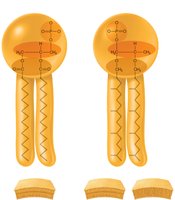
Cell Wall Structure and Function
Cell walls provide rigidity and protection. Bacteria use peptidoglycan; archaea use pseudopeptidoglycan or other polymers.
Peptidoglycan consists of glycan chains (NAG and NAM) cross-linked by peptides.
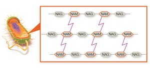
Gram Staining and Clinical Relevance
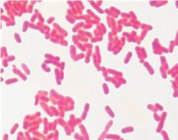
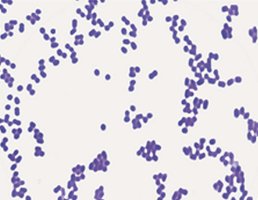
Gram staining differentiates bacteria based on cell wall structure.
Gram-positive: Thick peptidoglycan layer, stains purple, lacks outer membrane.
Gram-negative: Thin peptidoglycan layer, stains pink, has outer membrane with lipopolysaccharide (LPS).
Gram status affects susceptibility to antibiotics and disinfectants.
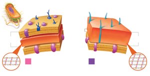
Comparing Gram-Negative and Gram-Positive Bacteria
Feature | Gram-Positive | Gram-Negative |
|---|---|---|
Peptidoglycan Layer | Thick | Thin |
Outer Membrane | Absent | Present |
Teichoic Acids | Present | Absent |
LPS | Absent | Present |
Stain Color | Purple | Pink |
Resistance | More sensitive to lysozyme | More resistant to chemicals |
Acid-Fast Staining
Acid-fast staining detects mycolic acid in cell walls. Acid-fast bacteria (e.g., Mycobacterium) appear red/pink and are slow-growing and resistant to drugs. 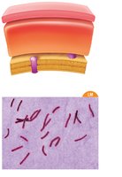
Mycoplasma and L-Forms
Mycoplasma species lack a cell wall and are pleomorphic.
L-forms are bacteria that have lost their cell wall, contributing to persistent infections.
Transport Across Cell Membranes
Passive Transport
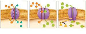
Diffusion: Movement of substances from high to low concentration.
Simple diffusion: Small, noncharged molecules and gases.
Facilitated diffusion: Uses membrane proteins for transport.
Osmosis
Osmosis is the diffusion of water across a selectively permeable membrane.
Isotonic: No net water movement.
Hypertonic: Water leaves cell, causing plasmolysis.
Hypotonic: Water enters cell, causing swelling or lysis.
Active Transport
Active transport requires energy and can move substances against a concentration gradient.
Primary active transport: Uses ATP (e.g., Na+-K+ pump).
Secondary active transport: Uses ion gradients (symporters and antiporters).
Phosphotransferase systems: Group translocation, phosphorylates transported substances.
External Structures for Adhesion, Movement, and Protection
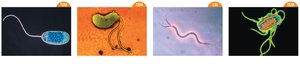
Flagella
Flagella are filamentous structures used for motility. They are built from flagellin and anchored by rings in the cell wall.
Gram-positive bacteria: Two rings.
Gram-negative bacteria: Four rings.

Movement is controlled by a chemotaxis system (run and tumble).
Types of movement: Chemotaxis (chemical), Phototaxis (light), Aerotaxis (oxygen).
Flagella arrangements: Monotrichous (single), Lophotrichous (cluster), Amphitrichous (both poles), Peritrichous (all over).
Periplasmic Flagella (Axial Filaments)
Periplasmic flagella are located between the plasma membrane and cell wall, allowing spirochetes to move in a corkscrew motion. 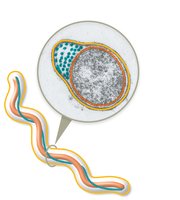
Fimbriae and Pili
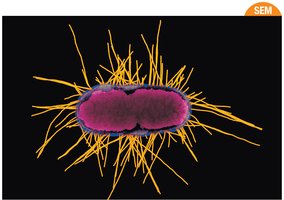
Fimbriae: Short, bristle-like structures for adhesion and biofilm formation.
Pili: Longer, more rigid structures for adhesion, motility, and gene transfer (conjugation).
Glycocalyx
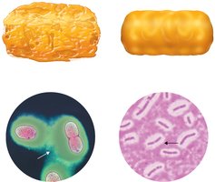
The glycocalyx is a carbohydrate-enriched layer that aids in adhesion and protection.
Slime layer: Unorganized and loosely associated.
Capsule: Well-organized and tightly associated.
Intracellular Structures
Cytoplasm and Nucleoid
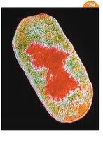
Cytoplasm: Site of most biochemical reactions.
Nucleoid region: Contains the single, circular chromosome.
Ribosomes
Prokaryotic ribosomes (70S) are composed of a large (50S) and small (30S) subunit. They synthesize proteins by linking amino acids. 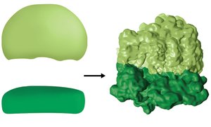
Cytoskeleton
Prokaryotic cytoskeletons are composed of protein filaments that provide structure and support.
Inclusion Bodies
Inclusion bodies are storage granules for substances such as carbon-fixing enzymes (carboxysomes) and magnetic iron (magnetosomes). 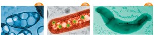
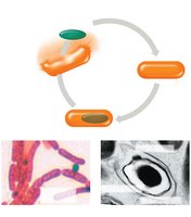
Endospores
Formation and Function
Endospores are metabolically inactive structures that allow certain bacteria to survive harsh conditions. They are highly resistant to environmental stresses.
When conditions improve, endospores germinate into vegetative cells.
Medically important genera: Bacillus, Clostridium, Clostridioides.
Clinical Case Example: Streptococcal Pharyngitis
Case Summary
A 5-year-old patient presented with symptoms of streptococcal pharyngitis. The causative agent, Streptococcus pyogenes, is a Gram-positive, nonmotile, encapsulated prokaryote.
Cell shape: Cocci, arranged in chains (streptococci).
Penicillin-family drugs are effective due to the presence of peptidoglycan in the cell wall.
No endospores or acid-fast properties expected.
Cell structures present: Capsule, cell wall, plasma membrane, nucleoid, ribosomes.
Absent structures: Flagella, endospores, acid-fast cell wall.
Not classified in domain Archaea.
Visual Summary