 뒤로
뒤로Introduction to Prokaryotic Cells: Structure, Shapes, and Cell Wall Differences
스터디 가이드 - 스마트 노트
 자료에 맞춘 맞춤형 노트, 핵심 정의, 예시, 맥락을 확장해 제공합니다.
Introduction to Prokaryotic Cells
Overview of Prokaryotic Cell Structure
Prokaryotic cells are unicellular organisms that lack a membrane-bound nucleus and organelles. They are structurally simpler than eukaryotic cells and include bacteria and archaea. Understanding their shapes, cell division, and cell wall composition is fundamental in microbiology.
Prokaryotic Cell Shapes
Diversity of Prokaryotic Shapes
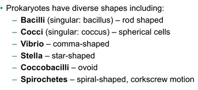
Prokaryotes exhibit a wide variety of cell shapes, which are important for classification and identification. The main shapes include:
Bacilli (rod-shaped)
Cocci (spherical cells)
Vibrio (comma-shaped)
Stella (star-shaped)
Coccobacilli (ovoid)
Spirochetes (spiral-shaped, corkscrew motion)
Arrangements and Additional Shapes
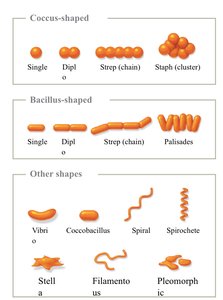
Prokaryotic cells can also be classified based on their arrangements and less common shapes:
Coccus-shaped arrangements: Single, Diplococcus (pairs), Streptococcus (chains), Staphylococcus (clusters)
Bacillus-shaped arrangements: Single, Diplobacillus (pairs), Streptobacillus (chains), Palisades (side-by-side)
Other shapes: Vibrio, Coccobacillus, Spiral, Spirochete, Stella, Filamentous, Pleomorphic
Surface Area to Volume Ratio in Cells
Importance of Surface Area to Volume Ratio
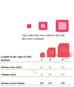
The surface area to volume ratio (SA:V) is a critical factor in cell biology. It influences the rate of nutrient uptake and waste elimination. Smaller cells have a higher SA:V, allowing for more efficient exchange with their environment, which is essential for survival and growth.
Formula for Surface Area of a Cube:
Formula for Volume of a Cube:
Surface Area to Volume Ratio:
As the size of the cell increases, the SA:V decreases, limiting the efficiency of transport processes.
Length of one edge of cube (inches) | Surface area (in2) | Volume (in3) | Surface area/volume |
|---|---|---|---|
2 | 24 | 8 | 3.0 |
4 | 96 | 64 | 1.5 |
6 | 216 | 216 | 1.0 |
Prokaryotic Cell Division
Binary Fission
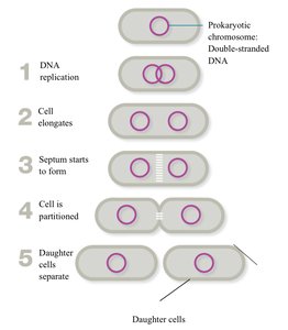
Prokaryotic cells reproduce asexually through a process called binary fission. This process ensures the rapid multiplication of prokaryotes under favorable conditions.
DNA replication: The circular chromosome is duplicated.
Cell elongation: The cell grows, and the chromosomes move to opposite ends.
Septum formation: A dividing wall (septum) begins to form in the center.
Partitioning: The septum completely divides the cell into two compartments.
Daughter cell separation: Two genetically identical daughter cells are formed.
Prokaryotic Cell Membranes
Differences Between Bacterial and Archaeal Membranes
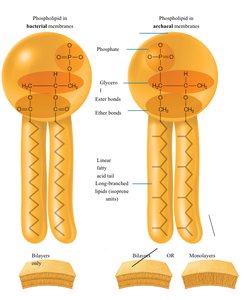
Prokaryotic cell membranes differ between bacteria and archaea, particularly in their lipid composition and bonding:
Bacterial membranes: Composed of phospholipids with ester bonds and unbranched fatty acid tails, forming bilayers.
Archaeal membranes: Composed of phospholipids with ether bonds and branched isoprene chains, which can form bilayers or monolayers. These adaptations provide stability in extreme environments.
Gram-Positive vs. Gram-Negative Cell Walls
Key Differences in Cell Wall Structure
Gram staining differentiates bacteria based on their cell wall structure, which affects their physical properties, susceptibility to antibiotics, and staining characteristics.
Cell Feature | Gram-Negative | Gram-Positive |
|---|---|---|
Outer membrane | Yes | No |
Lipid A (endotoxin) | Yes | No |
Porins | Yes | No |
Teichoic acids | No | Yes |
Peptidoglycan | Thin layer (10–20% of cell wall) | Thick layer (70–80% of cell wall) |
Gram staining color | Pink | Purple |
Resistant to physical disruption and drying? | No | Yes |
Susceptibility to anionic (negatively charged) detergents | Low | High |
Penicillin susceptibility | Low | High |
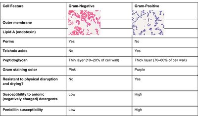
Example: Staphylococcus aureus is Gram-positive, while Escherichia coli is Gram-negative.
