 뒤로
뒤로Lab 1: Syllabus, Lab Safety, and Introduction to the Microscope
스터디 가이드 - 스마트 노트
 자료에 맞춘 맞춤형 노트, 핵심 정의, 예시, 맥락을 확장해 제공합니다.
Syllabus & Lab Safety
The Microscope
Parts and Handling of the Microscope
The compound light microscope is a fundamental tool in microbiology, allowing for the observation of microorganisms and cellular structures. Proper handling and identification of microscope parts are crucial for effective use and maintenance.
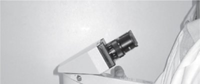
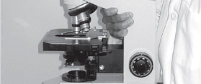
Major Parts: Ocular lenses (eyepieces), objective lenses, arm, base, stage, coarse and fine adjustment knobs, condenser, diaphragm, and illuminator.
Carrying the Microscope: Always carry the microscope with one hand on the arm and the other supporting the base to prevent damage.

Microscope Terminology
Resolution: The ability of a lens to distinguish two closely spaced points as separate entities.
Contrast: The difference in light intensity between the image and the adjacent background, essential for visualizing transparent specimens.
Types of Objective Lenses
Microscopes are equipped with several objective lenses of varying magnifications:
Scanning (4X): Used for initial specimen location and overview.
Low Power (10X): For general viewing and larger specimen details.
High-Dry (40X): For detailed observation of smaller structures.
Oil Immersion (100X): Used with immersion oil to achieve maximum resolution for observing bacteria and fine details.



Total Magnification
Total magnification is calculated by multiplying the magnification of the ocular lens by that of the objective lens:
Formula:
Example: If the ocular lens is 10X and the objective lens is 40X, then total magnification.
Using the Bright Field Microscope
Proper use of the microscope involves a series of steps to ensure clear visualization and prevent equipment damage:
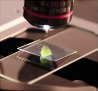
Rotate the objective lenses to the scanning lens (4X).
Raise the stage using the coarse focusing knob.
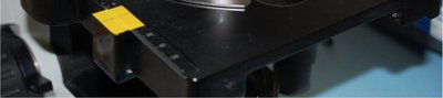
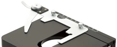
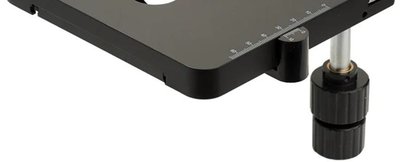
Secure the slide with the stage holder.
Use the mechanical stage knobs to position the slide so that light passes through the specimen.
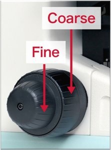
Focus on the specimen using the coarse focusing knob, then sharpen the image with the fine focusing knob.
Switch to higher magnification objectives as needed, using only the fine adjustment knob for focusing at higher powers.
Do not use the coarse adjustment knob with high-dry (40X) or oil immersion (100X) objectives to avoid damaging the slide or lens.





Oil Immersion Technique
The oil immersion lens (100X) is used to achieve the highest resolution. Immersion oil prevents light refraction, allowing more light to enter the lens and improving image clarity.
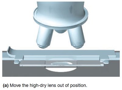
Move the high-dry lens out of position.
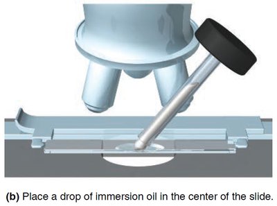
Place a drop of immersion oil on the slide.
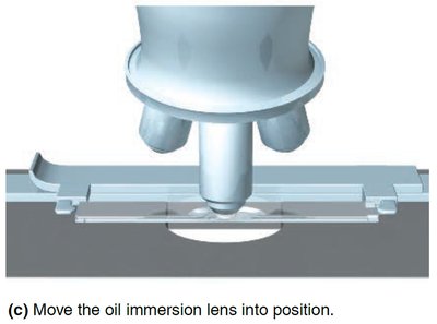
Rotate the oil immersion lens into position and use only the fine adjustment knob to focus.
Microscope Use and Care Tips
Adjust the aperture diaphragm to reduce chromatic aberration.
Use the condenser knob to concentrate the light beam on the specimen.
Position your eyes comfortably and adjust the ocular lenses for your pupillary distance and vision.
Always clean and return slides after use, and lower the stage with the 4X objective in place before storing the microscope.
Cell Morphology and Multicellular Arrangement
Introduction to Cell Morphology
Cell morphology refers to the shape and arrangement of microbial cells, which is critical for identification and classification in microbiology.
Common Shapes: Cocci (spherical), bacilli (rod-shaped), spirilla (spiral), and others.
Arrangements: Cells may occur singly, in pairs (diplo-), chains (strepto-), clusters (staphylo-), or other groupings.
Special Structures
Bacterial Capsule: A gelatinous outer layer that protects bacteria from desiccation and immune responses.
Endospores: Highly resistant, dormant structures formed by some bacteria for survival in harsh conditions.
Flagella and Motility: Flagella are whip-like appendages that enable bacterial movement.
Additional info: Understanding cell morphology and special structures is foundational for later topics such as microbial classification, pathogenesis, and laboratory identification techniques.
