 뒤로
뒤로Microbial Cell Structure and Function: The Cell Envelope
스터디 가이드 - 스마트 노트
 자료에 맞춘 맞춤형 노트, 핵심 정의, 예시, 맥락을 확장해 제공합니다.
The Cell Envelope
Overview of the Cell Envelope
The cell envelope is a series of layered structures that surround the cytoplasm of microbial cells, governing their interactions with the environment. It typically consists of the cytoplasmic membrane, cell wall, outer membrane, and sometimes S-layers. The composition and structure of the cell envelope vary significantly among Bacteria and Archaea, contributing to their physiological diversity and environmental adaptability.
2.1 The Cytoplasmic Membrane
Structure and Composition
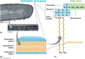
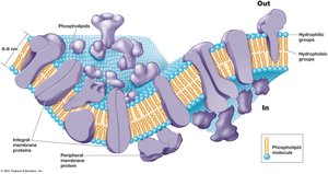
The cytoplasmic membrane surrounds the cytoplasm, separating it from the external environment. Its main function is selective permeability, allowing nutrients to enter and waste products to exit. The membrane is primarily composed of a phospholipid bilayer with embedded proteins, forming a fluid mosaic structure. The bilayer contains both hydrophobic (fatty acid tails) and hydrophilic (glycerol-phosphate head groups) components.
Hydrophobic region: Fatty acid tails face inward, away from water.
Hydrophilic region: Glycerol-phosphate head groups face outward, toward the cytoplasm or external environment.
Membrane proteins: Include integral (embedded), transmembrane (span the membrane), and peripheral (loosely attached) proteins.
Archaeal Cytoplasmic Membranes
Archaeal membranes differ from those of Bacteria and Eukarya:
Contain ether linkages in phospholipids (Bacteria/Eukarya have ester linkages).
Use isoprenoid chains instead of fatty acids.
Additional info: These differences contribute to the stability of archaeal membranes in extreme environments.
Functions of the Cytoplasmic Membrane
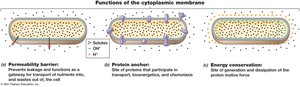
Permeability barrier: Prevents leakage and controls the passage of substances.
Protein anchor: Holds proteins involved in transport, bioenergetics, and chemotaxis.
Energy conservation: Site of generation and dissipation of the proton motive force, essential for ATP synthesis.
2.2 Transporting Nutrients into the Cell
Active Transport Mechanisms
Microbial cells use active transport to accumulate solutes against concentration gradients. Transporters are classified into three main mechanisms:
Simple transport: Uses a single transmembrane protein, driven by the proton motive force.
Group translocation: Involves a series of proteins; the transported molecule is chemically modified during passage.
ABC system (ATP-Binding Cassette): Consists of a binding protein, transmembrane transporter, and ATP-hydrolyzing protein; uses ATP for energy.
Simple transport can be:
Symport: Solute and H+ are cotransported in the same direction (e.g., E. coli lac permease).
Antiport: Solute and H+ are transported in opposite directions.
2.3 The Cell Wall
Function and Importance
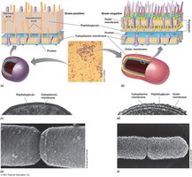
The cell wall provides structural strength, maintains cell shape, and prevents osmotic lysis. Most bacteria are classified as Gram-positive or Gram-negative based on their cell wall structure and Gram stain reaction.
Gram-Positive vs. Gram-Negative Cell Envelopes
Gram-positive: Thick peptidoglycan cell wall (20–35 nm), up to 90% peptidoglycan, often with teichoic acids.
Gram-negative: Thin peptidoglycan layer, outer membrane, and periplasmic space.
Bacterial Cell Walls: Peptidoglycan
Peptidoglycan: A rigid polysaccharide layer unique to Bacteria, providing strength and shape.
Stabilized by peptide cross-links; in Gram-positives, may include peptide interbridges (e.g., five glycines in Staphylococcus aureus).
Teichoic acids: Acidic molecules embedded in Gram-positive cell walls, sometimes covalently linked to membrane lipids (lipoteichoic acids).
Lysozyme: Enzyme that cleaves glycosidic bonds in peptidoglycan; found in human secretions as a defense mechanism.
Penicillin: Antibiotic that blocks formation of peptide cross-links in peptidoglycan.
Archaeal Cell Walls
Lack peptidoglycan and typically lack an outer membrane.
Most have an S-layer (protein shell) as the main wall component.
Some methanogens have a pseudomurein cell wall, similar to peptidoglycan but resistant to lysozyme and penicillin.
2.4 LPS: The Outer Membrane
Structure and Function of the Outer Membrane
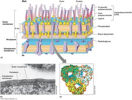
The outer membrane is a defining feature of Gram-negative bacteria, consisting of a lipid bilayer external to the cell wall. It contains lipopolysaccharide (LPS), which is important for surface recognition, virulence, and structural strength.
LPS: Composed of lipid A (toxic endotoxin), core polysaccharide, and O-polysaccharide.
Porins: Transmembrane proteins that allow the passage of solutes.
Periplasm: The space between the cytoplasmic and outer membranes, containing various enzymes and transport proteins.
2.5 Diversity of Cell Envelope Structure
S-Layers and Alternative Configurations
S-layers are paracrystalline protein or glycoprotein structures that, when present, form the outermost layer of the cell envelope. They provide strength, protection, shape, and facilitate cell surface interactions. Some Bacteria and Archaea lack cell walls but have tough cytoplasmic membranes, often containing sterols (e.g., Mycoplasmas in Bacteria, Thermoplasma in Archaea).
Some Archaea have only an S-layer outside the cytoplasmic membrane.
Others may have pseudomurein cell walls, with or without an S-layer.
Rarely, Archaea may possess an outer membrane.
Additional info: The diversity in cell envelope structure reflects adaptation to various environmental pressures and ecological niches.
