뒤로
뒤로Microscopy, Microbial Cultivation, and the Molecular Basis of Microbial Diversity
스터디 가이드 - 스마트 노트
 자료에 맞춘 맞춤형 노트, 핵심 정의, 예시, 맥락을 확장해 제공합니다.
Microscopy and the Origins of Microbiology
Light Microscopy and the Discovery of Microorganisms
Microbiology as a scientific discipline began with the invention and use of the microscope. Early pioneers such as Robert Hooke and Antoni van Leeuwenhoek made foundational discoveries by observing microorganisms for the first time. Hooke described the fruiting structures of molds and coined the term "cells," while van Leeuwenhoek was the first to describe bacteria using a simple light microscope.
Magnification: The ability to make an object appear larger.
Resolution: The ability to distinguish two adjacent objects as distinct and separate; for light microscopes, the limit is about 0.2 μm.
Types of light microscopy include: bright-field, phase-contrast, differential interference contrast, dark-field, and fluorescence microscopy.
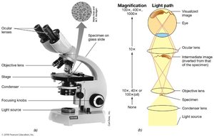
Compound light microscopes use two sets of lenses (objective and ocular) to form an image. The total magnification is the product of the magnifications of these lenses.
Bright-field microscopy visualizes specimens based on contrast differences between the specimen and its surroundings. Pigmented microbes can enhance contrast.
Formula:
Improving Contrast in Light Microscopy
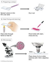
Contrast in light microscopy can be improved by staining, which involves the use of dyes that bind to specific cellular materials. Basic dyes, such as methylene blue, crystal violet, and safranin, are positively charged and bind strongly to negatively charged cell components. Simple stains use dried cells, while differential stains, such as the Gram stain, distinguish between different types of cells based on their cell wall structure.
Simple Stain: Uses a single dye to color cells, making them more visible under the microscope.
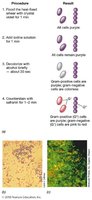
Differential Stain: Uses multiple dyes to differentiate between cell types (e.g., Gram-positive and Gram-negative bacteria).
Gram Stain: Gram-positive bacteria appear purple-violet, while Gram-negative bacteria appear pink due to differences in cell wall structure.
Probing Cell Structure: Electron Microscopy
Electron microscopy uses electrons instead of visible light to image cells and their structures, allowing for much higher resolution than light microscopy. Electromagnets serve as lenses, and the process operates in a vacuum. There are two main types:
Transmission Electron Microscopes (TEM): Used to view internal structures of cells.
Scanning Electron Microscopes (SEM): Used to view the surface details of cells and structures.
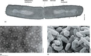
Microbial Cultivation Expands the Horizon of Microbiology
Pasteur and Spontaneous Generation
Louis Pasteur was instrumental in disproving the theory of spontaneous generation, which posited that life could arise spontaneously from nonliving material. Using the swan-necked flask experiment, Pasteur demonstrated that sterilized broth remained free of microbial growth unless exposed to microorganisms from the air. His work led to the development of sterilization methods, food preservation techniques, and vaccines for diseases such as anthrax, fowl cholera, and rabies.
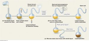
Koch, Infectious Disease, and Pure Cultures
Robert Koch established the link between specific microbes and infectious diseases, formulating the germ theory of disease. He identified the causative agents of anthrax, tuberculosis, and cholera, and developed solid media for obtaining pure cultures of microbes. Koch's postulates are a set of criteria used to prove that a specific microorganism causes a specific disease.
Pure cultures: Populations of cells derived from a single cell type.
Enrichment culture techniques: Methods to isolate microbes with specific metabolic characteristics from nature.
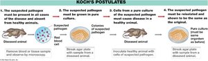
Discovery of Microbial Diversity
Sergei Winogradsky introduced the concept of chemolithotrophy, demonstrating that specific bacteria are involved in specific biogeochemical transformations, such as nitrogen and sulfur cycles. He also showed that some microbes can fix nitrogen and use carbon dioxide as a carbon source (autotrophy). This work expanded the focus of microbiology to include environmental and metabolic diversity.
Molecular Biology and the Unity and Diversity of Life
Molecular Basis of Life
Key discoveries in molecular biology established that DNA is the genetic material. Frederick Griffith demonstrated transformation in Streptococcus pneumoniae, while Watson, Crick, and Franklin elucidated the structure of DNA. These findings laid the foundation for understanding genetic transfer and heredity in microorganisms.
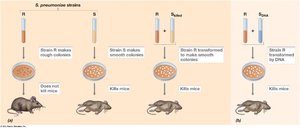
Woese and the Tree of Life
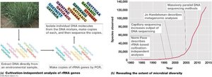
Carl Woese used ribosomal RNA (rRNA) sequences to infer evolutionary relationships among organisms, leading to the discovery of the domain Archaea and the construction of the three-domain tree of life (Bacteria, Archaea, Eukarya). This approach revealed that most microbial diversity had not yet been cultured and could be studied using cultivation-independent methods such as metagenomics.
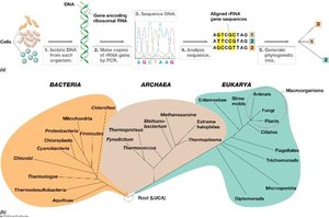
Analysis of Environmental rRNA Genes and Microbial Diversity
Advances in DNA sequencing technology have enabled the recovery and analysis of microbial genomes directly from environmental samples, greatly expanding our understanding of microbial diversity. Metagenomics allows for the study of microbial communities without the need for cultivation, revealing many previously unknown species.