 뒤로
뒤로Parasitology: Protozoans, Helminths, and Arthropods in Microbiology
스터디 가이드 - 스마트 노트
 자료에 맞춘 맞춤형 노트, 핵심 정의, 예시, 맥락을 확장해 제공합니다.
Parasitology: Overview
Cellular Living Organisms and Classification
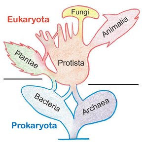
Parasitology is the study of parasites, their biology, and their impact on human health. Parasites include protozoans, helminths, and arthropods, which are classified based on their cellular structure and life cycles. The tree of life divides organisms into Prokaryota (Bacteria, Archaea) and Eukaryota (Fungi, Animalia, Plantae, Protista).
Protozoans: Characteristics and Examples
Protozoans are single-celled eukaryotes, many of which are found in freshwater environments. They lack a cell wall, possess ribosomes (60S/40S), and do not produce endospores but may form protective cysts. Their reproduction can be asexual or sexual, often involving complex life cycles. Infectious protozoans include Plasmodium (malaria), Trypanosoma (African sleeping sickness), Giardia (diarrhea), Entamoeba (diarrhea), Naegleria (CNS infection), Trichomonas (vaginal infection), and Toxoplasma (toxoplasmosis).
Locomotion in Protozoans
Protozoans move using specialized structures:
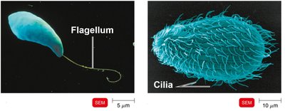
Cilia (e.g., Paramecium): Short, hair-like protrusions that propel organisms.
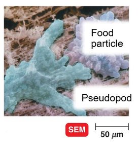
Pseudopodia (e.g., Amoeba): Cell extensions that flow in the direction of travel.
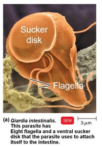
Flagella (e.g., Giardia): Longer, whip-like extensions of the cell.

Physical Forms and Life Cycle Stages
Protozoans exhibit different forms depending on environmental conditions and developmental stage:
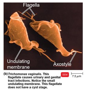
Trophozoite: Proliferative, infective form (e.g., Trichomonas vaginalis).
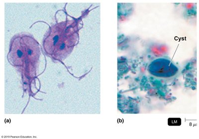
Cyst: Protective form, can survive outside the host for extended periods.
Reproduction
Sexual Reproduction: Conjugation (union of nuclei, seen in ciliates).
Asexual Reproduction: Binary fission (division into two cells), Schizogony (multiple fission).
Diseases Caused by Protozoans
Trichomoniasis
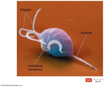
Caused by Trichomonas vaginalis, found in the vagina and male urinary tract. It lacks mitochondria and does not form cysts. Symptoms include discharge, itching, and painful urination, primarily in women. Humans are the only host.
Giardiasis
Caused by Giardia intestinalis (or G. lamblia), found in the small intestine of mammals. It lacks mitochondria and causes nausea, diarrhea, severe gas, cramping, and fatigue.

Toxoplasmosis
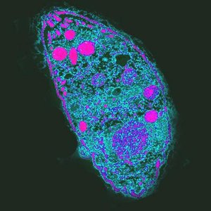
Caused by Toxoplasma gondii, an intracellular parasite. Trophozoites reproduce in cats, and oocysts are passed through cat feces. Infection is particularly risky for pregnant women.
Naegleria fowleri (Brain-eating Amoeba)
Found in freshwater, Naegleria fowleri enters the CNS via the nose and causes primary amebic meningoencephalitis (PAM). Symptoms include vomiting, headache, fever, coma, and death. Survival is rare.
Helminths: Parasitic Worms
General Characteristics
Helminths are multicellular eukaryotic animals with multiple organ systems. They reproduce sexually, producing eggs, and may require single or multiple hosts. Adult worms can be dioecious (separate sexes) or monoecious/hermaphroditic (both sexes in one individual).
Morphological Classification
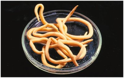
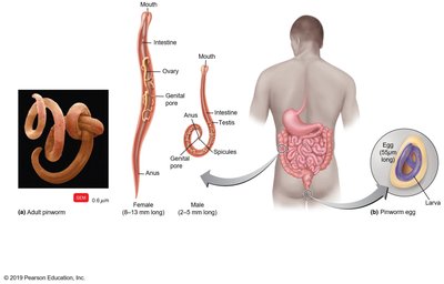
Nematodes: Round worms, cylindrical and tapered, complete digestive system.
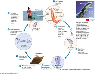
Trematodes: Flukes, flat and unsegmented.
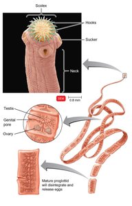
Cestodes: Tapeworms, flat and segmented.
Nematodes: Pinworm (Enterobius vermicularis)
Adults live in the large intestine. Females deposit eggs near the anus, which are spread via ingestion or contaminated linens.
Trematodes: Schistosoma mansoni
Flukes enter through the skin and travel in the circulatory system. Eggs can cause organ damage as they move through the bloodstream.
Cestodes: Taenia solium (Pork Tapeworm)
Tapeworms use suckers or hooks to attach to the host's intestinal mucosa. Eggs are passed through feces.
Parasitic Arthropods
Characteristics and Types
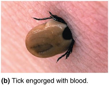
Arthropods are animals with external skeletons and segmented bodies. Many act as vectors, transmitting infectious microbes. They reproduce sexually and produce eggs.
Arachnida (8 legs): Spiders, mites, ticks. Examples: Lyme disease, babesiosis.
Crustacea (4 antennae): Crabs, crayfish.
Insecta (6 legs): Bees, flies, lice. Examples: Plague, malaria, Chagas disease, Dengue, Zika virus disease.
Summary Table: Classification of Parasitic Organisms
Type | Cellularity | Key Features | Examples |
|---|---|---|---|
Protozoans | Single-celled | No cell wall, complex life cycles, cyst formation | Plasmodium, Giardia, Trichomonas |
Helminths | Multicellular | Organ systems, sexual reproduction, eggs | Pinworm, Schistosoma, Taenia |
Arthropods | Multicellular | External skeleton, segmented body, vector role | Ticks, mosquitoes, lice |
Additional info: Academic context was added to clarify classification, life cycles, and disease relevance for exam preparation.
