Ch. 3 - Observing Microorganisms Through a Microscope
Tortora - Microbiology: An Introduction 14th Edition
Tortora14th EditionMicrobiology: An IntroductionISBN: 9780138200398당신이 사용하는 게 아니라요?교과서 변경
3장, 문제 9

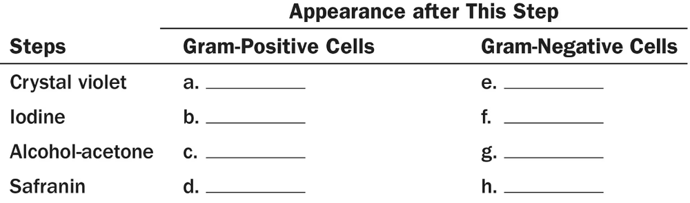
Fill in the following table regarding the Gram stain:
Table with steps of Gram stain and blank spaces to fill in the appearance of Gram-positive and Gram-negative cells after each step.

검증된 단계별 안내
1
Step 1: Understand the purpose of the Gram stain, which is to differentiate bacteria into Gram-positive and Gram-negative groups based on the structure of their cell walls.
Step 2: Identify the key components of the Gram stain procedure: crystal violet (primary stain), iodine (mordant), alcohol or acetone (decolorizer), and safranin (counterstain).
Step 3: For each step in the table, describe what happens to Gram-positive bacteria: they retain the crystal violet-iodine complex and appear purple because of their thick peptidoglycan layer.
Step 4: For each step, describe what happens to Gram-negative bacteria: they lose the crystal violet-iodine complex during decolorization due to their thinner peptidoglycan layer and outer membrane, and then take up the safranin counterstain, appearing pink/red.
Step 5: Fill in the table by matching each step of the Gram stain with the expected color and structural changes in both Gram-positive and Gram-negative bacteria.

비슷한 문제에 대한 검증된 영상 답변:

이 영상 해법은 위 문제에 도움이 된다고 튜터들이 추천한 것입니다.
영상 길이:
1m
도움이 되었나요?

주요 개념

질문에 올바르게 답하기 위해 반드시 이해해야 하는 핵심 개념들은 다음과 같습니다.

Gram Stain Procedure

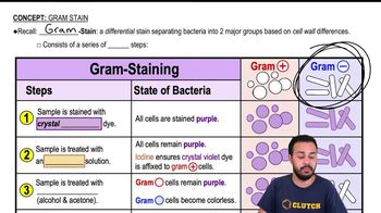
The Gram stain is a differential staining technique used to classify bacteria into Gram-positive or Gram-negative based on their cell wall properties. It involves four steps: crystal violet staining, iodine treatment, alcohol decolorization, and counterstaining with safranin. Each step affects how bacteria retain or lose the stain.
추천 영상:
04:57
Gram Stain

Bacterial Cell Wall Structure

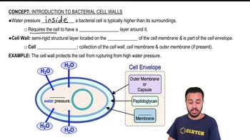
The difference in Gram stain results is due to variations in bacterial cell walls. Gram-positive bacteria have a thick peptidoglycan layer that retains the crystal violet-iodine complex, appearing purple. Gram-negative bacteria have a thin peptidoglycan layer and an outer membrane, causing them to lose the initial stain and take up the counterstain, appearing pink.
추천 영상:
03:44
Introduction to Bacterial Cell Walls

Interpretation of Gram Stain Results

Understanding how to interpret the colors and morphology after Gram staining is essential. Purple indicates Gram-positive bacteria, while pink indicates Gram-negative. This distinction helps in bacterial identification and guides antibiotic treatment decisions.
추천 영상:
04:57
Gram Stain