Ch. 3 - Observing Microorganisms Through a Microscope
Tortora - Microbiology: An Introduction 14th Edition
Tortora14th EditionMicrobiology: An IntroductionISBN: 9780138200398당신이 사용하는 게 아니라요?교과서 변경
3장, 문제 9

In 1996, scientists described a new tapeworm parasite that had killed at least one person. The initial examination of the patient’s abdominal mass was most likely made using:
a. Brightfield microscopy
b. Darkfield microscopy
c. Electron microscopy
d. Phase-contrast microscopy
e. Fluorescence microscopy

검증된 단계별 안내
1
Step 1: Understand the context of the problem. The question involves identifying the microscopy technique most likely used for the initial examination of a patient's abdominal mass containing a tapeworm parasite.
Step 2: Recall the characteristics of each microscopy type: brightfield microscopy uses visible light and is commonly used for stained or naturally pigmented specimens; darkfield microscopy enhances contrast in unstained samples; electron microscopy provides very high resolution but is more complex and used for ultrastructural details; phase-contrast microscopy enhances contrast in transparent specimens without staining; fluorescence microscopy uses fluorescent dyes to highlight specific structures.
Step 3: Consider the practical aspects of an initial clinical examination of a parasite in tissue. Typically, the first examination is done using a straightforward and widely available method that allows visualization of the parasite within tissue or fluid samples.
Step 4: Evaluate which microscopy technique fits this scenario best. Brightfield microscopy is the standard initial method for examining stained tissue sections or smears to identify parasites, as it is simple and effective for observing morphology.
Step 5: Conclude that the initial examination was most likely performed using brightfield microscopy, as it is the most common and practical method for initial parasite identification in clinical samples.

비슷한 문제에 대한 검증된 영상 답변:

이 영상 해법은 위 문제에 도움이 된다고 튜터들이 추천한 것입니다.
영상 길이:
1m
도움이 되었나요?

주요 개념

질문에 올바르게 답하기 위해 반드시 이해해야 하는 핵심 개념들은 다음과 같습니다.

Brightfield Microscopy

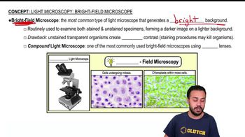
Brightfield microscopy is the most common light microscopy technique where the specimen is illuminated with white light and observed against a bright background. It is widely used for examining stained or naturally pigmented samples, including parasites in tissue sections or bodily fluids.
추천 영상:
03:46
Light Microscopy: Bright-Field Microscopes

Electron Microscopy

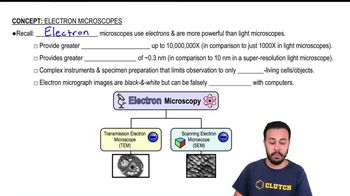
Electron microscopy uses a beam of electrons instead of light to achieve much higher resolution, allowing visualization of ultrastructural details of microorganisms. It is typically used for detailed structural analysis rather than initial clinical diagnosis due to complexity and cost.
추천 영상:
03:08
Electron Microscopes

Microscopy Techniques for Parasite Identification

Different microscopy methods serve specific purposes in parasite detection: brightfield is common for initial examination, darkfield enhances contrast of live, unstained organisms, phase-contrast improves visualization of transparent specimens, and fluorescence uses labeled antibodies or dyes for specific detection.
추천 영상:
03:46
Light Microscopy: Bright-Field Microscopes