 뒤로
뒤로NMR Spectroscopy: Principles and Applications in Organic Chemistry
스터디 가이드 - 스마트 노트
 자료에 맞춘 맞춤형 노트, 핵심 정의, 예시, 맥락을 확장해 제공합니다.
Nuclear Magnetic Resonance (NMR) Spectroscopy
Introduction to NMR Spectroscopy
NMR spectroscopy is a powerful analytical technique used to determine the structure of organic compounds by identifying the carbon–carbon and carbon–hydrogen framework. It relies on the magnetic properties of certain atomic nuclei, particularly those with an odd number of protons or neutrons, such as 1H, 13C, 15N, 19F, and 31P. Nuclei with even numbers of both protons and neutrons (e.g., 12C, 16O) are NMR inactive.
Spin States and Magnetic Field Effects
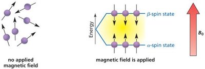
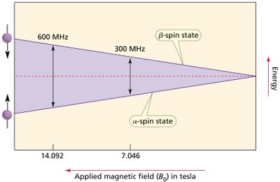
When a strong magnetic field is applied to a sample, the nuclei align either with or against the field, creating two distinct spin states: α (lower energy) and β (higher energy). The energy difference between these states depends on the strength of the applied magnetic field.
α-spin state: aligned with the magnetic field (lower energy)
β-spin state: aligned against the magnetic field (higher energy)
Operating Frequency and Spectrometer Function
The operating frequency of an NMR spectrometer is directly related to the strength of the magnetic field. Higher magnetic fields result in greater energy differences and higher resonance frequencies.
Key Equation:
The energy difference () between spin states is given by:
: applied magnetic field
: Planck’s constant
: gyromagnetic ratio
Fourier Transform NMR (FT-NMR)
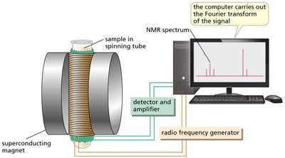
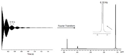
Modern NMR spectrometers use pulsed Fourier transform methods, where a constant magnetic field is applied and a short radiofrequency (rf) pulse excites all nuclei simultaneously. The resulting signal, called Free Induction Decay (FID), is mathematically converted into an NMR spectrum using a Fourier transform.
Proton NMR: Chemical Shifts and Signal Interpretation
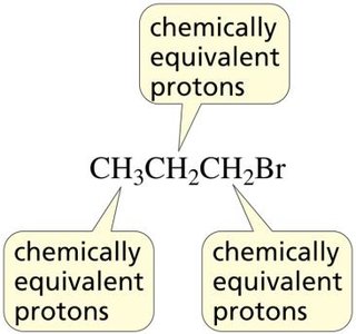
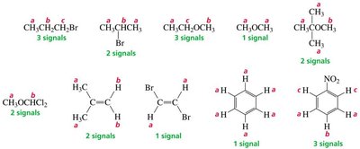
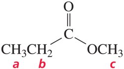
Protons in different chemical environments experience different local magnetic fields due to surrounding electrons, resulting in distinct NMR signals. The number of signals corresponds to the number of sets of chemically equivalent protons.
Chemically equivalent protons: protons in the same environment give the same signal.
Chemical shift (δ): indicates the environment around the nucleus, measured in parts per million (ppm).
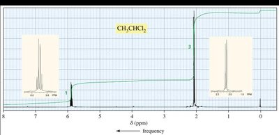
Peak integration: area under the peak is proportional to the number of protons.
Signal splitting: reveals information about adjacent protons.
The Reference Compound: Tetramethylsilane (TMS)
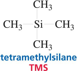
TMS is used as the reference compound in NMR spectroscopy, appearing at 0 ppm. All chemical shifts are measured relative to TMS.
Interpreting Chemical Shifts
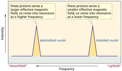
The chemical shift depends on the electronic environment. Electron-withdrawing groups deshield protons, causing signals to appear at higher frequencies (downfield). Electron-donating groups shield protons, resulting in lower frequency signals (upfield).
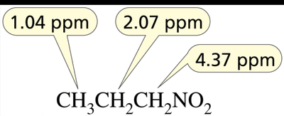
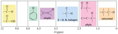
Table: Chemical Shift Values for Common Proton Types
Type of Proton | Approximate δ (ppm) |
|---|---|
Methyl (CH3) | 0.85 |
Methylene (CH2) | 1.20 |
Methine (CH) | 1.55 |
Allylic (C=C–CH3) | 1.7 |
Acetyl (CO–CH3) | 2.1 |
Aromatic (benzene ring) | 6.5–8 |
Aldehyde (CHO) | 9.0–10 |
Carboxylic acid (COOH) | 10–12 |
Signal Integration and Splitting
The area under each NMR signal is proportional to the number of protons responsible for that signal. Signal splitting (multiplicity) occurs due to interactions with neighboring protons, following the (N + 1) rule:
Singlet: N = 0
Doublet: N = 1
Triplet: N = 2
Quartet: N = 3
Multiplet: N > 3 or unequal coupling constants
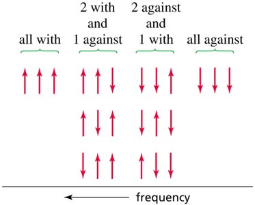
Coupling Constants (J)
The coupling constant (J) is the distance between adjacent peaks in a split NMR signal, measured in hertz (Hz). Coupled protons have the same J value, and the magnitude of J can distinguish between cis and trans isomers in alkenes.
Table: Typical Coupling Constants
Type of Coupling | J (Hz) |
|---|---|
Geminal (same carbon) | 12 |
Vicinal (adjacent carbons) | 7 |
Long-range (four bonds) | 1 |
Trans vinylic | 15 |
Cis vinylic | 10 |
Special Cases: Enantiotopic and Diastereotopic Hydrogens
Enantiotopic hydrogens are chemically equivalent and not distinguishable by NMR. Diastereotopic hydrogens are not chemically equivalent and react differently, showing distinct NMR signals.
Carbon-13 NMR Spectroscopy
Carbon-13 NMR provides information about the carbon framework of organic molecules. The number of signals reflects the number of different types of carbons. Chemical shifts range over 220 ppm, and TMS is used as the reference compound.
Table: Chemical Shift Values for Common Carbon Types
Type of Carbon | Approximate δ (ppm) |
|---|---|
Methyl (R–CH3) | 0–35 |
Methylene (R–CH2–R) | 15–55 |
Methine (R3–CH) | 25–55 |
Alkyne (C≡C) | 70–90 |
Aromatic (benzene ring) | 110–170 |
Carbonyl (C=O) | 165–220 |
Advanced NMR Techniques
Two-dimensional NMR (e.g., COSY, HETCOR) provides information about coupling between protons and between protons and carbons, allowing for more detailed structural analysis.
NMR in Medical Imaging: MRI
Magnetic Resonance Imaging (MRI) uses NMR principles to visualize internal structures of the body. Different tissues yield distinct signals, which are processed to generate cross-sectional images.
Summary of NMR Spectroscopy
The number of signals indicates the number of sets of equivalent protons or carbons.
Chemical shifts reveal the nature of the chemical environment.
Integration values show the relative number of protons.
Splitting patterns provide information about neighboring protons.
Coupling constants identify coupled protons and distinguish isomers.
Example Applications
Structure elucidation: Determining the connectivity and environment of atoms in organic molecules.
Isomer identification: Distinguishing between cis/trans and other stereoisomers.
Medical imaging: MRI for non-invasive visualization of tissues.
